Difference between revisions of "File:IPLab8HSVEncephalitis5.jpg"
Seung Park (talk | contribs) (This is a high-power photomicrograph of the previous section. At this power it is easier to see the blood vessel with the perivascular hemorrhage and mild perivascular lymphocytic cuffing (1). In addition, the areas of edema and loss of neurophil (2) c...) |
(No difference)
|
Latest revision as of 02:35, 21 August 2013
This is a high-power photomicrograph of the previous section. At this power it is easier to see the blood vessel with the perivascular hemorrhage and mild perivascular lymphocytic cuffing (1). In addition, the areas of edema and loss of neurophil (2) can be better appreciated. Red shrunken neurons and glia with pyknotic nuclei (3) are also evident at this power.
File history
Click on a date/time to view the file as it appeared at that time.
| Date/Time | Thumbnail | Dimensions | User | Comment | |
|---|---|---|---|---|---|
| current | 02:35, 21 August 2013 | 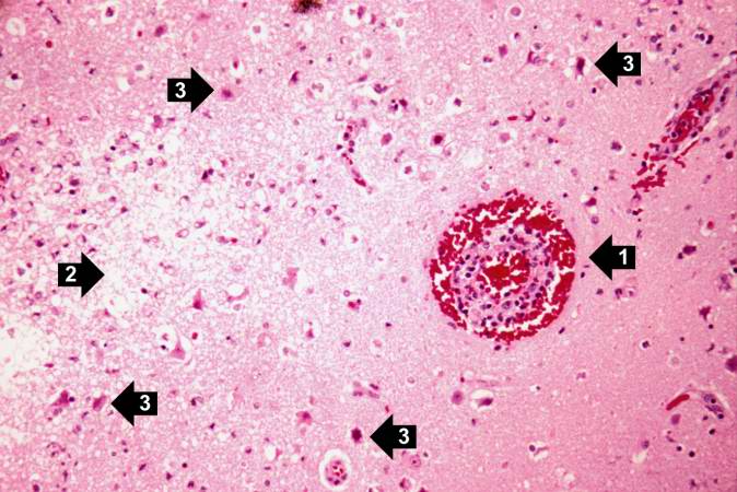 | 674 × 450 (62 KB) | Seung Park (talk | contribs) | This is a high-power photomicrograph of the previous section. At this power it is easier to see the blood vessel with the perivascular hemorrhage and mild perivascular lymphocytic cuffing (1). In addition, the areas of edema and loss of neurophil (2) c... |
- You cannot overwrite this file.
File usage
The following page links to this file: