Difference between revisions of "File:IPLab8HSVEncephalitis7.jpg"
Seung Park (talk | contribs) (This is a high-power photomicrograph demonstrating clear areas, which indicate edema, and numerous shrunken red necrotic cells (1). At this power, it can be seen that eosinophilic intranuclear inclusion bodies have displaced chromatin to the periphery ...) |
(No difference)
|
Latest revision as of 02:36, 21 August 2013
This is a high-power photomicrograph demonstrating clear areas, which indicate edema, and numerous shrunken red necrotic cells (1). At this power, it can be seen that eosinophilic intranuclear inclusion bodies have displaced chromatin to the periphery of the nucleus in some cells (2).
File history
Click on a date/time to view the file as it appeared at that time.
| Date/Time | Thumbnail | Dimensions | User | Comment | |
|---|---|---|---|---|---|
| current | 02:36, 21 August 2013 | 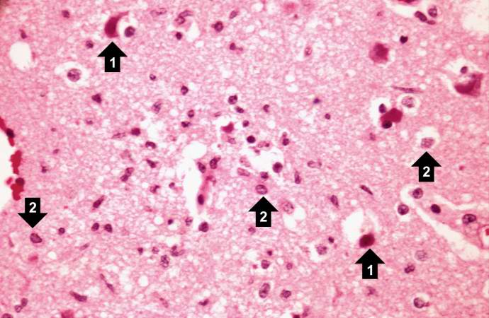 | 690 × 450 (55 KB) | Seung Park (talk | contribs) | This is a high-power photomicrograph demonstrating clear areas, which indicate edema, and numerous shrunken red necrotic cells (1). At this power, it can be seen that eosinophilic intranuclear inclusion bodies have displaced chromatin to the periphery ... |
- You cannot overwrite this file.
File usage
The following page links to this file: