Difference between revisions of "File:IPLab8HSVEncephalitis10.jpg"
Seung Park (talk | contribs) (This is another high-power photomicrograph of a cell containing an intranuclear inclusion body (arrow). Note that the nuclear chromatin has been pushed to the outer edges of the nucleus.) |
(No difference)
|
Latest revision as of 02:37, 21 August 2013
This is another high-power photomicrograph of a cell containing an intranuclear inclusion body (arrow). Note that the nuclear chromatin has been pushed to the outer edges of the nucleus.
File history
Click on a date/time to view the file as it appeared at that time.
| Date/Time | Thumbnail | Dimensions | User | Comment | |
|---|---|---|---|---|---|
| current | 02:37, 21 August 2013 | 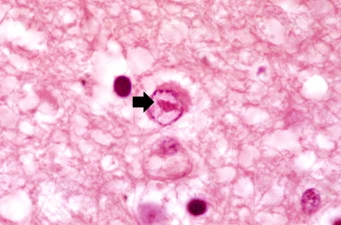 | 680 × 450 (40 KB) | Seung Park (talk | contribs) | This is another high-power photomicrograph of a cell containing an intranuclear inclusion body (arrow). Note that the nuclear chromatin has been pushed to the outer edges of the nucleus. |
- You cannot overwrite this file.
File usage
The following page links to this file: