Difference between revisions of "File:IPLab8HBV8.jpg"
Seung Park (talk | contribs) (This higher-power photomicrograph of the previous section shows more clearly the HBsAg positive cells (arrows). Upon staining with H&E, these same cells exhibit a "ground glass" appearance, which is due to the accumulation of HBsAg in the hepatocyte cy...) |
(No difference)
|
Latest revision as of 02:49, 21 August 2013
This higher-power photomicrograph of the previous section shows more clearly the HBsAg positive cells (arrows). Upon staining with H&E, these same cells exhibit a "ground glass" appearance, which is due to the accumulation of HBsAg in the hepatocyte cytoplasm.
File history
Click on a date/time to view the file as it appeared at that time.
| Date/Time | Thumbnail | Dimensions | User | Comment | |
|---|---|---|---|---|---|
| current | 02:49, 21 August 2013 | 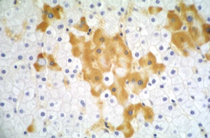 | 684 × 450 (37 KB) | Seung Park (talk | contribs) | This higher-power photomicrograph of the previous section shows more clearly the HBsAg positive cells (arrows). Upon staining with H&E, these same cells exhibit a "ground glass" appearance, which is due to the accumulation of HBsAg in the hepatocyte cy... |
- You cannot overwrite this file.
File usage
The following page links to this file: