Difference between revisions of "File:IPLab9RMSF8.jpg"
Seung Park (talk | contribs) (This is a high-power photomicrograph of a thrombosed vessel in the dermis. Note that the endothelial cells are missing along part of the circumference of the vessel (arrows)--this is where the main part of the thrombus has attached. Also note the infla...) |
(No difference)
|
Latest revision as of 03:43, 21 August 2013
This is a high-power photomicrograph of a thrombosed vessel in the dermis. Note that the endothelial cells are missing along part of the circumference of the vessel (arrows)--this is where the main part of the thrombus has attached. Also note the inflammation surrounding the vessel.
A thrombus is a solid mass resulting from the aggregation of blood constituents within the vascular system.
File history
Click on a date/time to view the file as it appeared at that time.
| Date/Time | Thumbnail | Dimensions | User | Comment | |
|---|---|---|---|---|---|
| current | 03:43, 21 August 2013 | 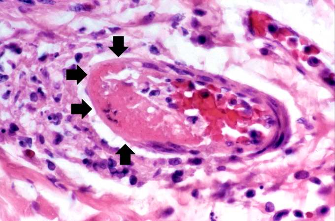 | 680 × 450 (49 KB) | Seung Park (talk | contribs) | This is a high-power photomicrograph of a thrombosed vessel in the dermis. Note that the endothelial cells are missing along part of the circumference of the vessel (arrows)--this is where the main part of the thrombus has attached. Also note the infla... |
- You cannot overwrite this file.
File usage
The following page links to this file: