Difference between revisions of "IPLab:Lab 9:Actinomycosis"
Seung Park (talk | contribs) |
Seung Park (talk | contribs) |
||
| (7 intermediate revisions by the same user not shown) | |||
| Line 13: | Line 13: | ||
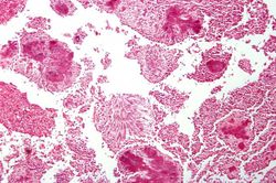
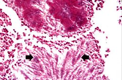
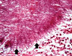
File:IPLab9Actinomycosis5.jpg|This is a high-power photomicrograph of an actinomycotic colony. The filamentous nature (arrows) of the actinomyces organisms is more easily appreciated at this power. | File:IPLab9Actinomycosis5.jpg|This is a high-power photomicrograph of an actinomycotic colony. The filamentous nature (arrows) of the actinomyces organisms is more easily appreciated at this power. | ||
</gallery> | </gallery> | ||
| + | |||
| + | == Virtual Microscopy == | ||
| + | <peir-vm>IPLab9Actinomycosis</peir-vm> | ||
| + | |||
| + | == Study Questions == | ||
| + | * <spoiler text="These organisms are ubiquitous in the environment but they seldom cause disease. Why?">Actinomyces requires an anaerobic environment deep within tissue to flourish. As a non-invasive bacterium, the Actinomyces organisms must be introduced into these deep tissues before causing harm.</spoiler> | ||
| + | |||
| + | == Additional Resources == | ||
| + | === Reference === | ||
| + | * [http://emedicine.medscape.com/article/211587-overview eMedicine Medical Library: Actinomycosis] | ||
| + | * [http://emedicine.medscape.com/article/960759-overview eMedicine Medical Library: Pediatric Actinomycosis] | ||
| + | * [http://www.merckmanuals.com/professional/infectious_diseases/anaerobic_bacteria/actinomycosis.html Merck Manual: Actinomycosis] | ||
| + | |||
| + | === Journal Articles === | ||
| + | * Yildiz O, Doganay M. [http://www.ncbi.nlm.nih.gov/pubmed/16582679 Actinomycoses and Nocardia pulmonary infections]. ''Curr Opin Pulm Med'' 2006 May;12(3):228-34. | ||
| + | |||
| + | === Images === | ||
| + | * [{{SERVER}}/library/index.php?/tags/2154-actinomycosis PEIR Digital Library: Actinomycosis Images] | ||
{{IPLab 9}} | {{IPLab 9}} | ||
[[Category: IPLab:Lab 9]] | [[Category: IPLab:Lab 9]] | ||
Latest revision as of 16:32, 3 January 2014
Contents
Clinical Summary[edit]
This 18-year-old black female felt well until one year before death, when she developed a persistent, progressive skin rash and weight loss. One month before death, draining abscesses appeared in the perirectal region. Biopsy showed actinomycosis. Despite treatment, the patient died.
Autopsy Findings[edit]
Autopsy revealed a large abscess around the cecum which had ruptured. The perirectal abscesses had originated from extensions of this pericecal abscess.
Images[edit]
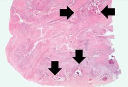
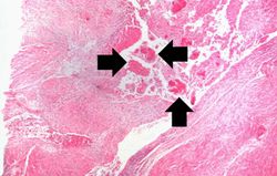
This is a low-power photomicrograph of the retroperitoneal abscess. At this magnification, multiple dark-staining foci can be appreciated. These foci are Actinomyces colonies (arrows). These colonies are known as "sulfur granules" because in gross specimens they are visible to the naked eye as yellow grains, thus resembling grains of sulfur.
Virtual Microscopy[edit]
Study Questions[edit]
Additional Resources[edit]
Reference[edit]
- eMedicine Medical Library: Actinomycosis
- eMedicine Medical Library: Pediatric Actinomycosis
- Merck Manual: Actinomycosis
Journal Articles[edit]
- Yildiz O, Doganay M. Actinomycoses and Nocardia pulmonary infections. Curr Opin Pulm Med 2006 May;12(3):228-34.
Images[edit]
| |||||
An abscess is a collection of pus (white blood cells) within a cavity formed by disintegrated tissue.
An abscess is a collection of pus (white blood cells) within a cavity formed by disintegrated tissue.
