Difference between revisions of "IPLab:Lab 3:Sarcoidosis"
Seung Park (talk | contribs) (→Journal Articles) |
(→Virtual Microscopy) |
||
| (6 intermediate revisions by one other user not shown) | |||
| Line 12: | Line 12: | ||
</gallery> | </gallery> | ||
| − | + | == Virtual Microscopy == | |
| + | === Lymph Node: Sarcoidosis === | ||
| + | <peir-vm>IPLab3Sarcoidosis</peir-vm> | ||
| + | === Lymph Node === | ||
| + | <peir-vm>UAB-Histology-00035</peir-vm> | ||
| + | |||
| + | == Study Questions == | ||
* <spoiler text="What is sarcoidosis?">The exact cause of sarcoidosis is unknown. Sarcoidosis is characterized by non-caseating granulomas in many tissues and organs.</spoiler> | * <spoiler text="What is sarcoidosis?">The exact cause of sarcoidosis is unknown. Sarcoidosis is characterized by non-caseating granulomas in many tissues and organs.</spoiler> | ||
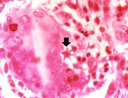
* <spoiler text="What is the characteristic phenotype of sarcoid granulomas?">The typical characteristic of a sarcoid granuloma is a non-caseating granuloma which consists of an aggregate of tightly-clustered epitheliod cells, often with Langhans’ or multinucleated giant cells. Occasionally, asteroid bodies may be seen.</spoiler> | * <spoiler text="What is the characteristic phenotype of sarcoid granulomas?">The typical characteristic of a sarcoid granuloma is a non-caseating granuloma which consists of an aggregate of tightly-clustered epitheliod cells, often with Langhans’ or multinucleated giant cells. Occasionally, asteroid bodies may be seen.</spoiler> | ||
| Line 31: | Line 37: | ||
=== Images === | === Images === | ||
| + | * [{{SERVER}}/library/index.php?/tags/531-sarcoidosis PEIR Digital Library: Sarcoidosis Images] | ||
| + | * [http://library.med.utah.edu/WebPath/LUNGHTML/LUNGIDX.html#2 WebPath: Granulomatous Disease] | ||
| − | + | == Related IPLab Cases == | |
| − | + | * [[IPLab:Lab 2:Metastatic Calcification|Lab 2: Lung: Metastatic Calcification]] | |
| − | |||
{{IPLab 3}} | {{IPLab 3}} | ||
[[Category: IPLab:Lab 3]] | [[Category: IPLab:Lab 3]] | ||
Latest revision as of 19:27, 17 September 2015
Contents
Clinical Summary[edit]
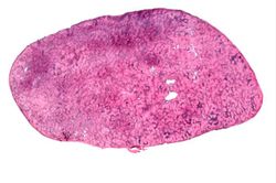
This 33-year-old white female was admitted for evaluation of abnormal findings on a chest x-ray. She was asymptomatic and a physical examination revealed no significant abnormalities. Laboratory results indicated hypercalcemia and elevated gamma globulin. Radiographic examination showed enlarged subcarinal, hilar, and right paratracheal lymph nodes. A right paratracheal lymph node was biopsied. Special stains for acid-fast bacilli and fungi were negative and a diagnosis of sarcoidosis was made.
Images[edit]
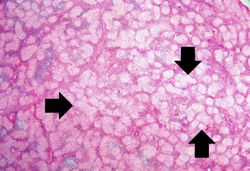
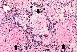
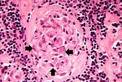
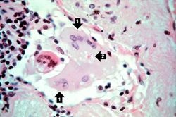
This is a photomicrograph of the small nodules (arrows) seen in the previous image. Close examination reveals that they are composed of large macrophages (epithelioid macrophages). These small granulomas form multiple series of reaction centers throughout the lymph node. Note the remaining lymphocytes surrounding the granulomas.
Virtual Microscopy[edit]
Lymph Node: Sarcoidosis[edit]
Lymph Node[edit]
Study Questions[edit]
Additional Resources[edit]
Reference[edit]
- eMedicine Medical Library: Acute Complications of Sarcoidosis
- eMedicine Medical Library: Sarcoidosis
- eMedicine Medical Library: Dermatologic Manifestations of Sarcoidosis
- eMedicine Medical Library: Neurosarcoidosis
- Merck Manual: Sarcoidosis
Journal Articles[edit]
- English JC, Patel PJ, Greer KE. Sarcoidosis. J Am Acad Dermatol 2001;44:725-43
- Johns CJ, Michele TM. The clinical management of sarcoidosis. A 50-year experience at the Johns Hopkins Hospital. Medicine (Baltimore) 1999 Mar;78(2):65-111.
Images[edit]
Related IPLab Cases[edit]
Hypercalcemia is the state of having increased levels of calcium in the blood.
