Difference between revisions of "IPLab:Lab 7:Malignant Melanoma"
Seung Park (talk | contribs) (Created page with "== Images == <gallery heights="250px" widths="250px"> File:IPLab7Melanoma1.jpg|This is a gross photograph of skin with melanoma. Note the black pigment, multiple satellite nod...") |
Seung Park (talk | contribs) |
||
| Line 1: | Line 1: | ||
| + | == Clinical Summary == | ||
| + | This 68-year-old white male had a local excision of a pigmented lesion (melanoma) on the skin of his back. Three years later he became aware of a "lump" in his left axilla. Examination confirmed the presence of a 2.3-cm nodular lesion. Subsequently, the patient underwent a surgical procedure for removal of axillary lymph nodes. | ||
| + | |||
== Images == | == Images == | ||
<gallery heights="250px" widths="250px"> | <gallery heights="250px" widths="250px"> | ||
Revision as of 14:34, 21 August 2013
Clinical Summary[edit]
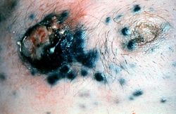
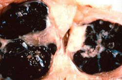
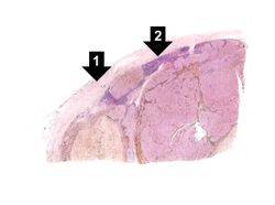
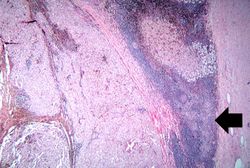
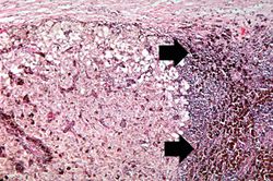
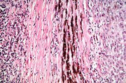
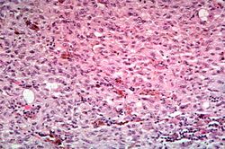
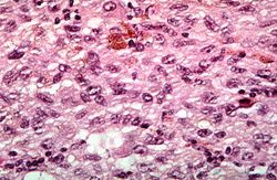
This 68-year-old white male had a local excision of a pigmented lesion (melanoma) on the skin of his back. Three years later he became aware of a "lump" in his left axilla. Examination confirmed the presence of a 2.3-cm nodular lesion. Subsequently, the patient underwent a surgical procedure for removal of axillary lymph nodes.
Images[edit]
Nodular hyperplasia of the prostate--characterized by large discrete prostatic nodules--is a common disorder in men over 50 years of age. The nodules cause the prostate to be enlarged and to have an increased weight. The human prostate is surrounded by a restrictive capsule. These nodules cause increased pressure within the capsule which leads to constriction of the urethra as it passes through the prostate. Urethral constriction leads to retention of urine.
