Clinical Summary[edit]
This 55-year-old white male had a long history of emphysema and a 60-70 pack-year smoking history. He was in his usual state of health until about one month before admission, at which time he developed increasing dyspnea on exertion. At the same time, his sputum increased from two tablespoons to half a cup of yellow blood-streaked sputum a day. Chest x-ray showed a right hilar mass. Sputum cytology revealed abnormal cells that were "positive for malignancy." He later developed pneumonia and fever. The patient expired soon thereafter.
Autopsy Findings[edit]
Significant findings included advanced carcinoma of the right main stem bronchus with extension across the carina to produce obstruction of the left main stem bronchus. There was left lower lobe pneumonia and left upper lobe atelectasis. Extensive metastases were present in regional lymph nodes as well as the pericardium, left atrium, and right kidney.
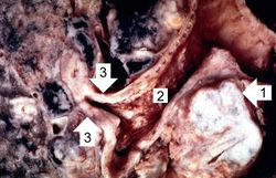
This is a gross photograph of bronchogenic carcinoma. The large tumor mass can be seen adjacent to the bronchus (1). Note that the epithelial surface of the bronchus is rough and irregular (2). The first branch off the right main stem bronchus is partially occluded by the thickened mucosa and submucosa (3).
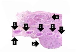
This is a low-power photomicrograph of bronchus showing normal mucosa (1) with transition to carcinoma (2). Note the bronchial cartilage (3) and the invasion of tumor through the entire wall of the bronchus with tumor extending to the serosal surface (4).
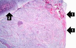
This is a photomicrograph of bronchus with ulcerated mucosal surface on the right (1). The submucosa is completely filled with tumor down to the cartilage (2).
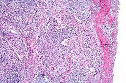
This is a higher-power photomicrograph of bronchus with the ulcerated mucosal surface on the right and tumor underneath.
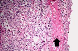
This is a higher-power photomicrograph of the mucosal surface (right) with an area of hemorrhage (arrow) and underlying tumor (left).
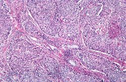
This is a photomicrograph of tumor from an area of invasion with compression of fibrous stroma and focal necrosis.
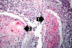
This is a high-power photomicrograph showing cytologic detail of the tumor with an area of necrosis (1) and a more differentiated area with keratin pearl formation (2).
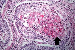
This is a high power photomicrograph of tumor with an area of central necrosis (arrow).
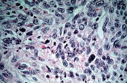
This high-power photomicrograph of tumor shows the cytologic detail of a less-differentiated area of neoplasm with cellular anaplasia.
Study Questions[edit]
Squamous cell carcinoma, and adenocarcinoma occur at approximately equal frequency, 25 to 40% each; small cell carcinoma, 20 to 25%; and large cell carcinoma, 10 to 15%.
- There is a positive relationship between tobacco smoking and lung cancer. Compared with nonsmokers, cigarette smokers have a ten-fold greater risk of developing lung cancer, and heavy smokers (more than 40 cigarettes per day for several years) have at least a 20-fold greater risk. Eighty per cent of lung cancers occur in smokers.
- Industrial hazards (uranium, asbestos, etc.).
- Air pollution (radon).
- Genetic factors. Occasional familial clustering has been observed suggesting a genetic component to lung cancer. Dominant oncogenes (c-myc in small cell carcinoma & K-ras in adenocarcinomas) and loss or inactivation of recessive tumor suppressor genes (p53 & retinoblastoma) have been reported in lung cancer.
- Scarring. Some lung cancers arise in the area of old scars and are termed scar carcinomas.
- Antidiuretic hormone (ADH), inducing hyponatremia due to inappropriate ADH secretion;
- adrenocorticotropic hormone (ACTH), producing Cushing’s syndrome;
- parathormone, parathyroid hormone-related peptide, prostaglandin E, and some cytokines, all implicated in the hypercalcemia often seen with lung cancer;
- calcitonin, causing hypocalcemia;
- gonadotropins, causing gynecomastia; and
- serotonin, associated with the carcinoid syndrome. The incidence of clinically significant syndromes related to these factors ranges from 1 to 10% of all lung cancer patients. Any one of the histologic types of tumors may occasionally produce any one of the hormones, but tumors producing ACTH and ADH are predominantly small cell carcinomas and those producing hypercalcemia are mostly squamous cell tumors.
The clinical course is somewhat variable but in general the prognosis is poor with a 5 year survival of approximately 9%.
