File list
This special page shows all uploaded files.
| Date | Name | Thumbnail | Size | User | Description | Versions |
|---|---|---|---|---|---|---|
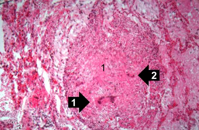
| 03:17, 19 August 2013 | IPLab3LobarPneumonia4.jpg (file) |  |
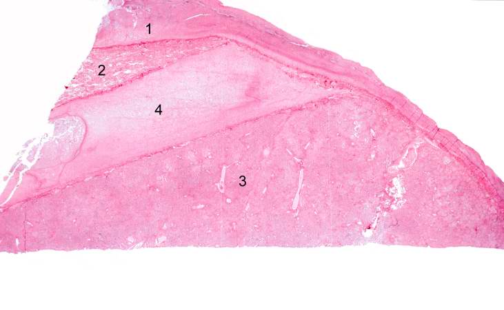
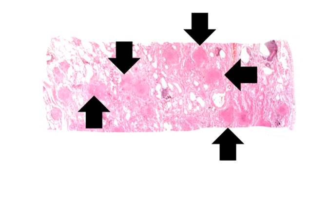
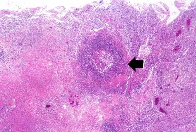
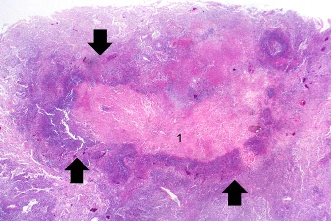
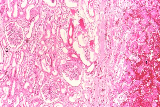
31 KB | Seung Park | This low-power photomicrograph of the lung shows: (1) markedly thickened pleura, indicating an inflammatory process which has been present for several days; (2) a small wedge-shaped segment of normal lung which is somewhat compressed due to artifact; (... | 1 |
| 03:17, 19 August 2013 | IPLab3LobarPneumonia3.jpg (file) |  |
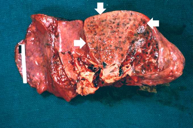
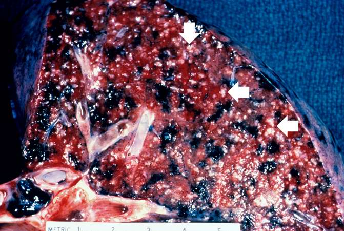
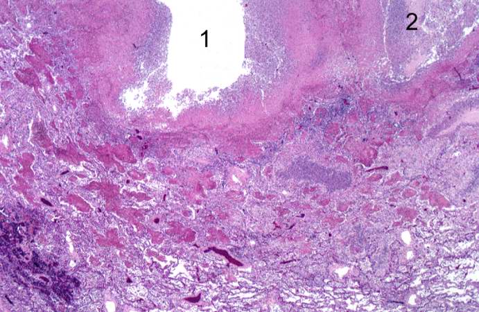
55 KB | Seung Park | This is a gross photograph of the right lung from the patient in this case. This lung shows complete consolidation with a marked infiltration of neutrophils throughout the tissue giving the lung a whitish discoloration. Note the extensive black pigment... | 1 |
| 03:16, 19 August 2013 | IPLab3LobarPneumonia2.jpg (file) |  |
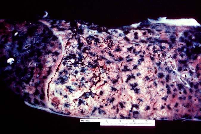
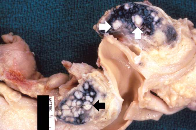
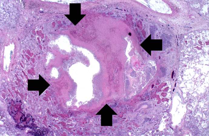
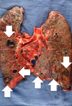
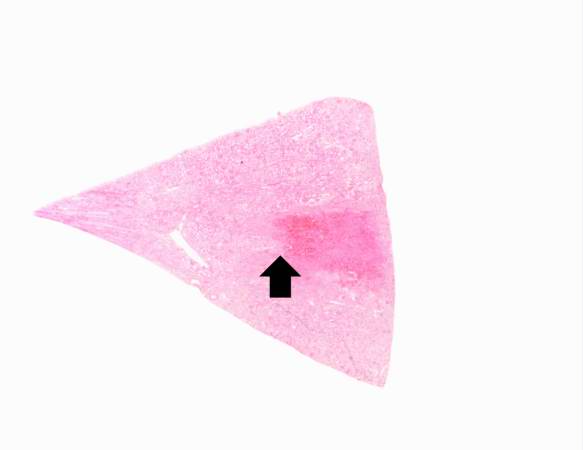
55 KB | Seung Park | This is a cut section of a lung from the preceding image. Note the whitish discoloration of the lung tissue in the upper lobe (arrows) compared to the normal collapsed and pink staining lung lobe in the left-hand portion of the photograph. The white di... | 1 |
| 03:15, 19 August 2013 | IPLab3LobarPneumonia1.jpg (file) |  |
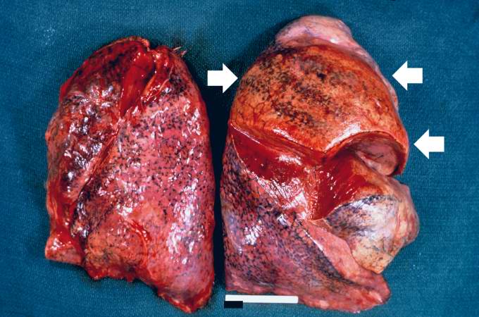
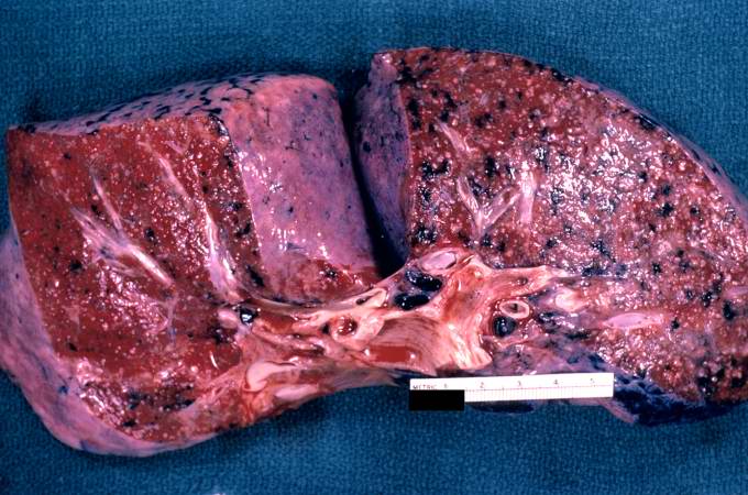
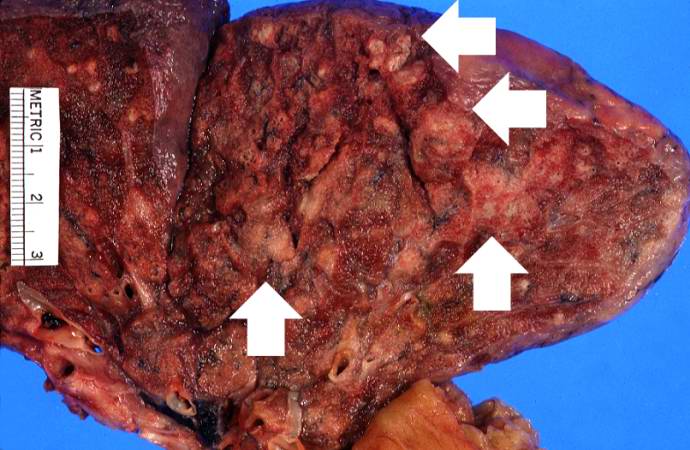
58 KB | Seung Park | This is a gross photograph of the lungs from a patient (not the patient from this case) with acute lobar pneumonia. The lung lobe in the upper-right portion of the photograph is affected with pneumonia (arrows). It has a whitish discoloration and appea... | 1 |
| 02:13, 19 August 2013 | IPLab3AcuteAppendicitis8.jpg (file) |  |
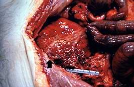
58 KB | Seung Park | This is a gross photograph of another example of peritonitis. Again note the fibrinosuppurative exudate covering the abdominal organs (arrows). | 1 |
| 02:12, 19 August 2013 | IPLab3AcuteAppendicitis7.jpg (file) |  |
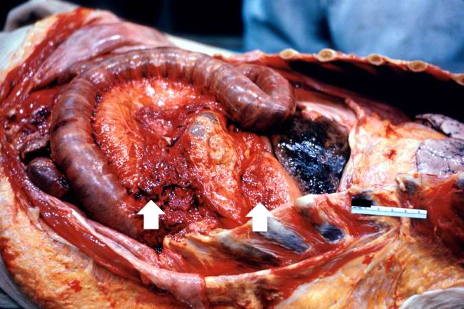
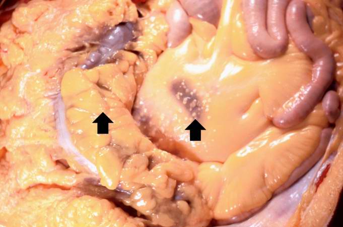
13 KB | Seung Park | This is a gross photograph of the open abdominal cavity of a patient with acute appendicitis. In this patient, there had been rupture of the appendix with spillage of intestinal contents into the abdominal cavity. This spillage resulted in an acute abd... | 1 |
| 02:12, 19 August 2013 | IPLab3AcuteAppendicitis6.jpg (file) |  |
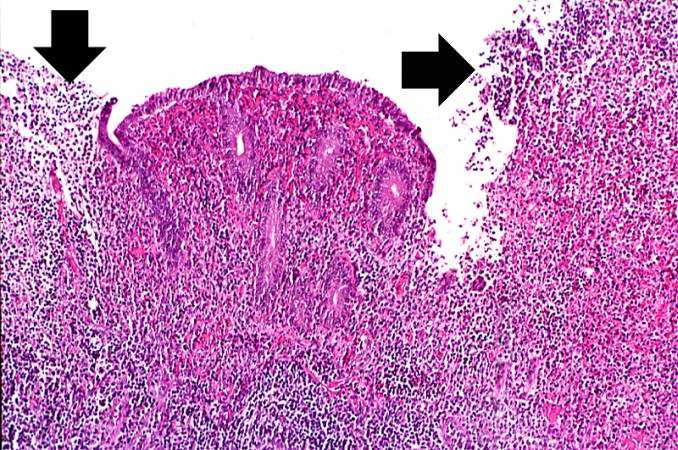
106 KB | Seung Park | This higher-power photomicrograph of the mucosal surface shows the loss of normal mucosal epithelium (arrows) and the inflammatory infiltrate. The principal inflammatory cell in this case of acute appendicitis is the neutrophil. | 1 |
| 02:12, 19 August 2013 | IPLab3AcuteAppendicitis5.jpg (file) |  |
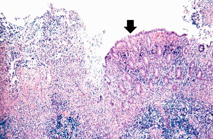
88 KB | Seung Park | This photomicrograph of the mucosal surface shows a small area with normal mucosal epithelium (arrow). This area is surrounded by areas of ulceration with an inflammatory infiltrate of lymphocytes and neutrophils. | 1 |
| 02:11, 19 August 2013 | IPLab3AcuteAppendicitis4.jpg (file) |  |
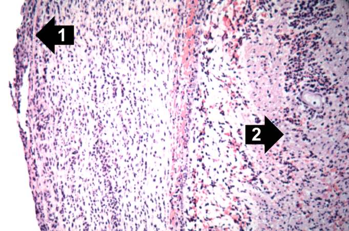
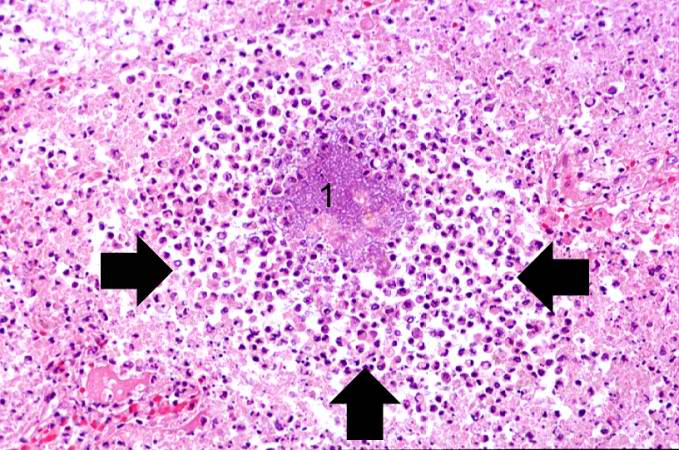
71 KB | Seung Park | This is a photomicrograph of the serosal surface of the appendix on the left (1) and the submucosal tissue in the center (2) with remnants of a lymphoid nodule. Surrounding this lymphoid nodule are masses of leukocytes which should not be present in a ... | 1 |
| 02:10, 19 August 2013 | IPLab3AcuteAppendicitis3.jpg (file) |  |
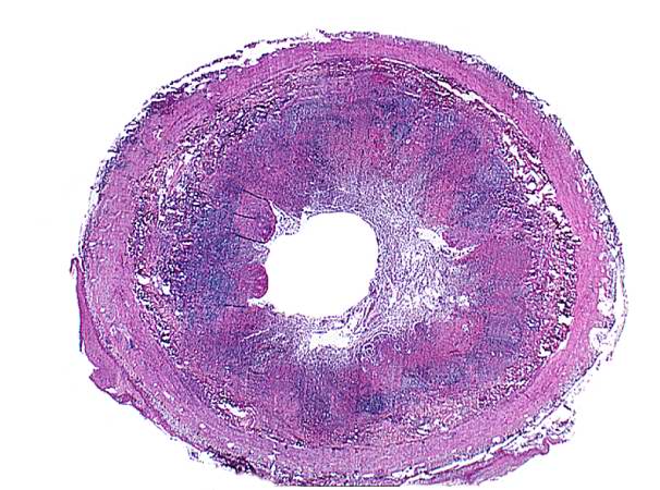
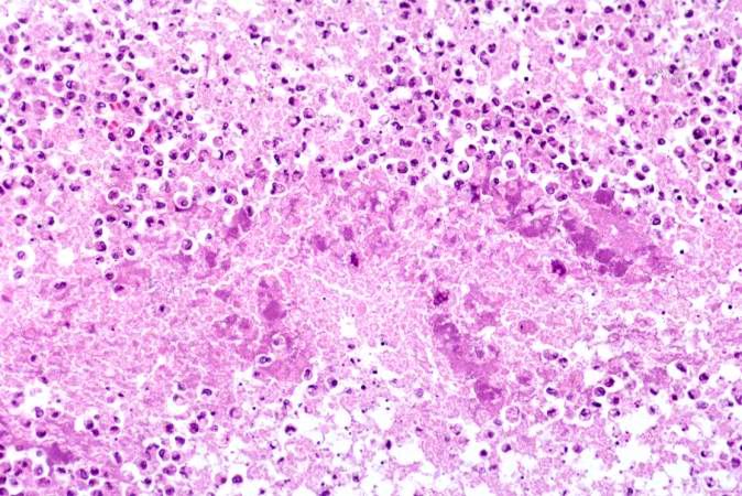
64 KB | Seung Park | This is a photomicrograph of an appendix exhibiting acute inflammation. Note that there are only remnants of mucosal tissue identifiable along the luminal border of this specimen. There is an extensive infiltration of leukocytes in this tissue which ca... | 1 |
| 02:10, 19 August 2013 | IPLab3AcuteAppendicitis2.jpg (file) |  |
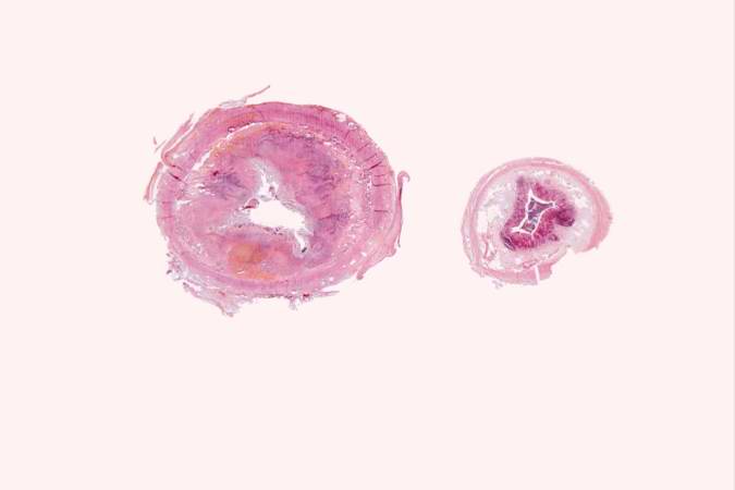
13 KB | Seung Park | This is a low-power photomicrograph of a normal appendix on the right and an appendix with acute inflammatory response on the left. Note the abundant blue-stained lymphoid tissue beneath the mucosal layer and the absence of blue-staining cells in the s... | 1 |
| 02:10, 19 August 2013 | IPLab3AcuteAppendicitis1.jpg (file) |  |
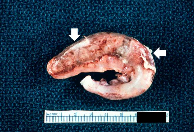
61 KB | Seung Park | This is a gross photograph of the appendix which was removed from this patient with acute appendicitis. Note the rough, shaggy material (arrows) on the surface due to deposition of fibrin and inflammatory cells. | 1 |
| 23:30, 18 August 2013 | IPLab2Hypertrophy6.jpg (file) |  |
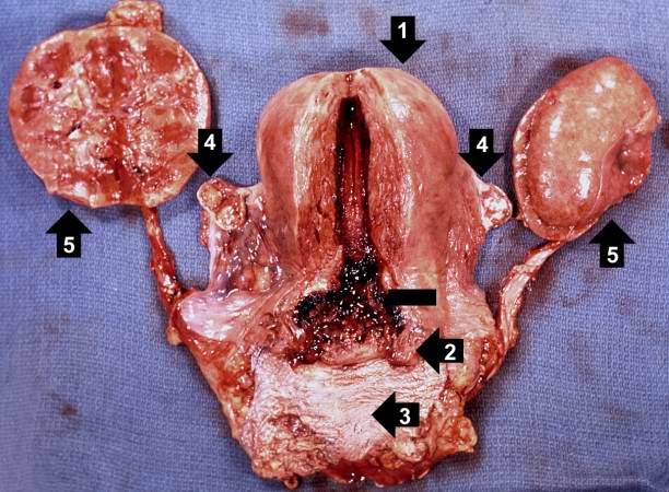
63 KB | Seung Park | This gross photograph shows an example of normal physiologic hypertrophy. The organs shown are an open uterus (1), cervix (2) and vagina (3), both ovaries (4) and both kidneys (5) from a woman who died shortly after normal delivery from causes unrelate... | 1 |
| 23:29, 18 August 2013 | IPLab2Hypertrophy5.jpg (file) |  |
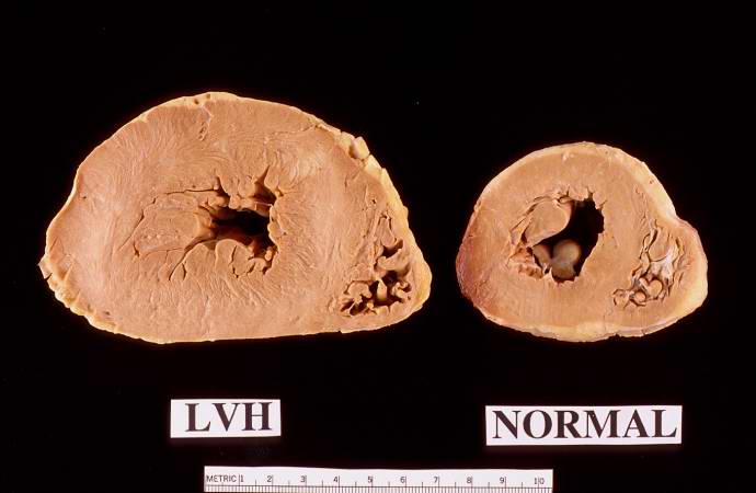
47 KB | Seung Park | This autopsy specimen was taken from another patient who had cardiac hypertrophy and congestive heart failure that resulted in dilation of the cardiac chambers. This heart was markedly enlarged (700 grams) but the congestive failure leads to dilation o... | 1 |
| 23:29, 18 August 2013 | IPLab2Hypertrophy1.jpg (file) |  |
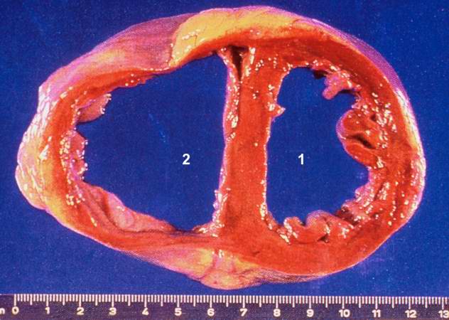
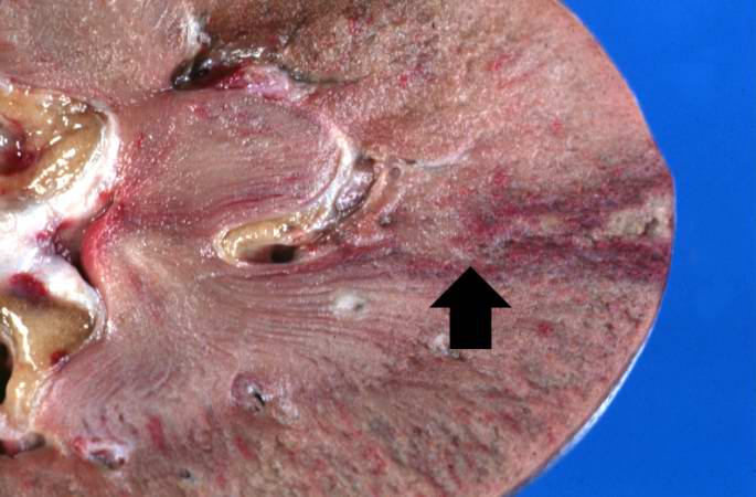
30 KB | Seung Park | This is a gross photograph of a cross section of a normal human heart taken at autopsy (right) and the heart from this case, which demonstrates concentric hypertrophy of the left ventricular wall. Note the marked thickening of the left ventricular wall... | 1 |
| 03:42, 16 August 2013 | IPLab1Prostate6.jpg (file) |  |
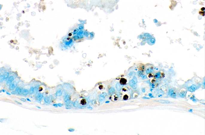
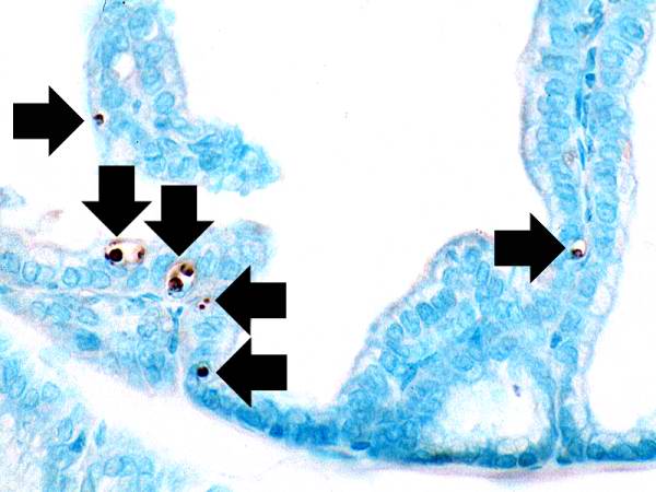
27 KB | Seung Park | This is a higher-power photomicrograph of prostatic epithelium with the TUNEL staining. Note the apoptotic cells (brown nuclei) in the epithelium as well as those floating freely. | 1 |
| 03:42, 16 August 2013 | IPLab1Prostate5.jpg (file) |  |
35 KB | Seung Park | This photomicrograph of prostatic epithelium demonstrates an in situ immunohistochemical technique that is used to identify the DNA fragments characteristic of apoptotic nuclei. This technique, terminal deoxynucleotidyl transferase-mediated dUTP-biotin... | 1 |
| 03:41, 16 August 2013 | IPLab1Prostate4.jpg (file) |  |
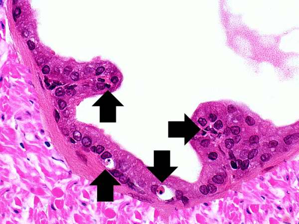
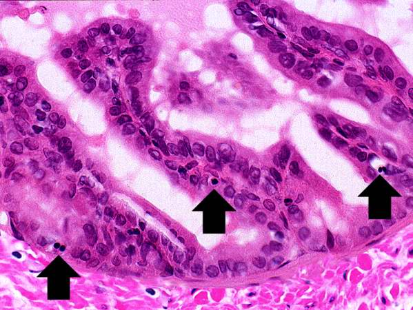
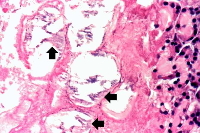
46 KB | Seung Park | Still another high-power photomicrograph of the prostatic epithelium demonstrates cells with pyknotic and fragmented nuclei (arrows). Again note the condensed and hypereosinophilic cytoplasm. | 1 |
| 03:41, 16 August 2013 | IPLab1Prostate3.jpg (file) |  |
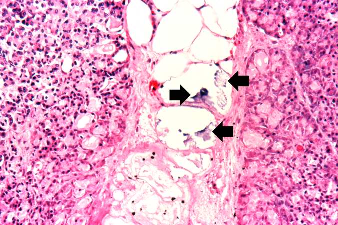
60 KB | Seung Park | Another high-power photomicrograph of the prostatic epithelium shows cells with pyknotic and fragmented nuclei (arrows). Note that the cytoplasm is condensed and hypereosinophilic. | 1 |
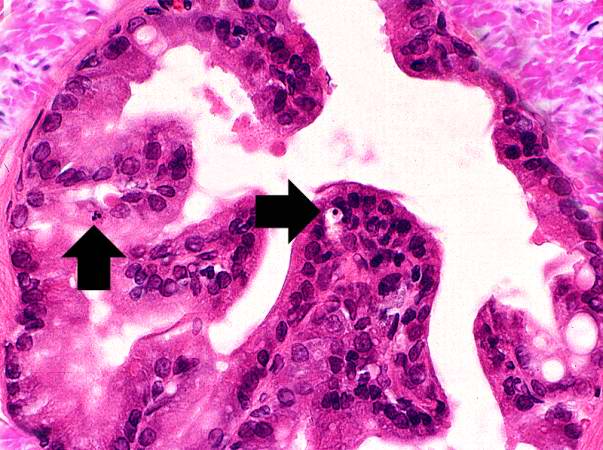
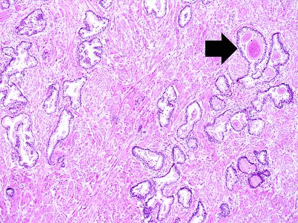
| 03:40, 16 August 2013 | IPLab1Prostate2.jpg (file) |  |
61 KB | Seung Park | 1 | |
| 03:40, 16 August 2013 | IPLab1Prostate1.jpg (file) |  |
86 KB | Seung Park | 1 | |
| 02:50, 16 August 2013 | IPLab1Tuberculosis7.jpg (file) |  |
64 KB | Seung Park | 1 | |
| 02:50, 16 August 2013 | IPLab1Tuberculosis6.jpg (file) |  |
64 KB | Seung Park | 1 | |
| 02:50, 16 August 2013 | IPLab1Tuberculosis5.jpg (file) |  |
58 KB | Seung Park | 1 | |
| 02:50, 16 August 2013 | IPLab1Tuberculosis4.jpg (file) |  |
21 KB | Seung Park | 1 | |
| 02:49, 16 August 2013 | IPLab1Tuberculosis3.jpg (file) |  |
42 KB | Seung Park | 1 | |
| 02:49, 16 August 2013 | IPLab1Tuberculosis2.jpg (file) |  |
72 KB | Seung Park | 1 | |
| 02:49, 16 August 2013 | IPLab1Tuberculosis1.jpg (file) |  |
66 KB | Seung Park | 1 | |
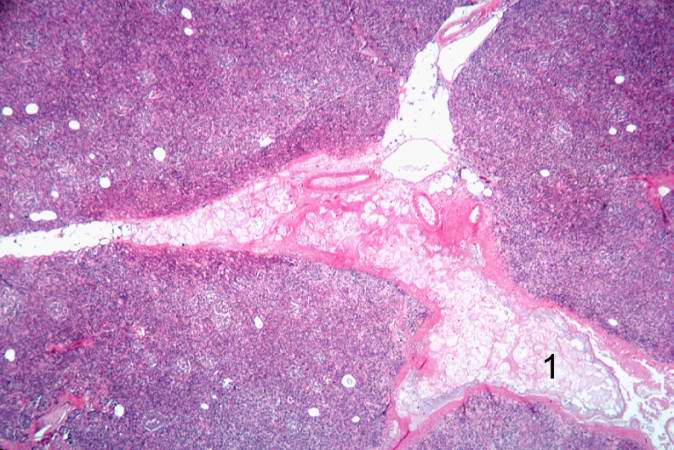
| 01:19, 16 August 2013 | IPLab1FatNecrosis9.jpg (file) |  |
52 KB | Seung Park | 1 | |
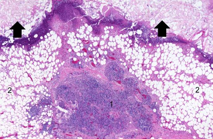
| 01:18, 16 August 2013 | IPLab1FatNecrosis8.jpg (file) |  |
72 KB | Seung Park | 1 | |
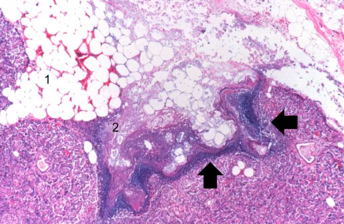
| 01:18, 16 August 2013 | IPLab1FatNecrosis7.jpg (file) |  |
81 KB | Seung Park | 1 | |
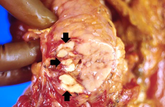
| 01:18, 16 August 2013 | IPLab1FatNecrosis6.jpg (file) |  |
83 KB | Seung Park | 1 | |
| 01:18, 16 August 2013 | IPLab1FatNecrosis5.jpg (file) |  |
68 KB | Seung Park | 1 | |
| 01:18, 16 August 2013 | IPLab1FatNecrosis4.jpg (file) |  |
76 KB | Seung Park | 1 | |
| 01:18, 16 August 2013 | IPLab1FatNecrosis3.jpg (file) |  |
65 KB | Seung Park | 1 | |
| 01:17, 16 August 2013 | IPLab1FatNecrosis2.jpg (file) |  |
42 KB | Seung Park | 1 | |
| 01:17, 16 August 2013 | IPLab1FatNecrosis1.jpg (file) |  |
36 KB | Seung Park | 1 | |
| 16:15, 15 August 2013 | IPLab1LungAbscess8.jpg (file) |  |
85 KB | Seung Park | 1 | |
| 16:15, 15 August 2013 | IPLab1LungAbscess7.jpg (file) |  |
79 KB | Seung Park | 1 | |
| 16:15, 15 August 2013 | IPLab1LungAbscess6.jpg (file) |  |
66 KB | Seung Park | 1 | |
| 16:15, 15 August 2013 | IPLab1LungAbscess5.jpg (file) |  |
56 KB | Seung Park | 1 | |
| 16:15, 15 August 2013 | IPLab1LungAbscess4.jpg (file) |  |
75 KB | Seung Park | 1 | |
| 16:15, 15 August 2013 | IPLab1LungAbscess3.jpg (file) |  |
62 KB | Seung Park | 1 | |
| 16:15, 15 August 2013 | IPLab1LungAbscess2.jpg (file) |  |
57 KB | Seung Park | 1 | |
| 16:14, 15 August 2013 | IPLab1LungAbscess1.jpg (file) |  |
30 KB | Seung Park | 1 | |
| 15:12, 15 August 2013 | IPLab1KidneyInfarction7.jpg (file) |  |
43 KB | Seung Park | 1 | |
| 15:12, 15 August 2013 | IPLab1KidneyInfarction6.jpg (file) |  |
64 KB | Seung Park | 1 | |
| 15:12, 15 August 2013 | IPLab1KidneyInfarction5.jpg (file) |  |
64 KB | Seung Park | 1 | |
| 15:12, 15 August 2013 | IPLab1KidneyInfarction4.jpg (file) |  |
71 KB | Seung Park | 1 | |
| 15:12, 15 August 2013 | IPLab1KidneyInfarction3.jpg (file) |  |
11 KB | Seung Park | 1 |