File list
This special page shows all uploaded files.
| Date | Name | Thumbnail | Size | User | Description | Versions |
|---|---|---|---|---|---|---|
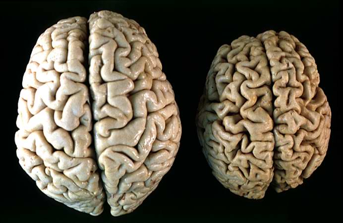
| 16:12, 19 August 2013 | IPLab2Atrophy10.jpg (file) |  |
43 KB | Peter Anderson | This gross photograph shows a normal brain (left) and a brain from a geriatric patient (right). Note the decreased size, the narrowed gyri, and the widened sulci of the brain from this octogenarian. What is the cause of atrophy in this case? | 1 |
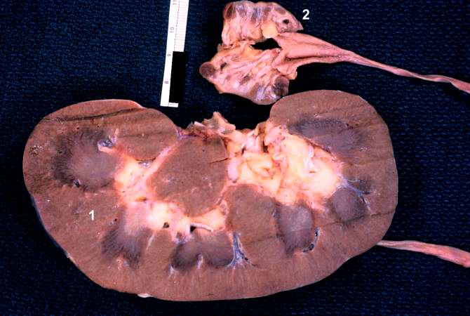
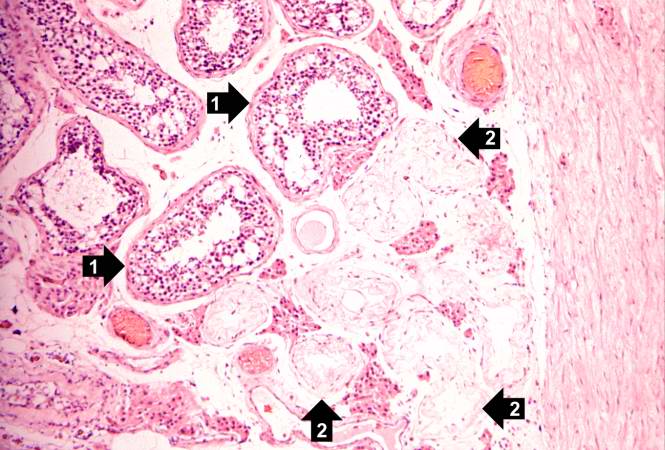
| 16:11, 19 August 2013 | IPLab2Atrophy9.jpg (file) |  |
47 KB | Peter Anderson | The two kidneys in this slide are from the same patient. One kidney (1) is relatively normal, although increased in size due to compensatory hypertrophy. The other kidney (2) is very small with only rudimentary nodules of renal parenchyma. This kidney ... | 1 |
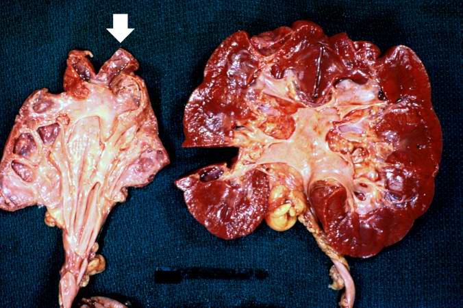
| 16:11, 19 August 2013 | IPLab2Atrophy8.jpg (file) |  |
64 KB | Peter Anderson | These kidneys were removed from a patient who had blockage of one ureter leading to increased pressure in the renal pelvis. The increased pressure produced hydronephrosis (arrow) in one kidney. What is the cause of atrophy in this case? | 1 |
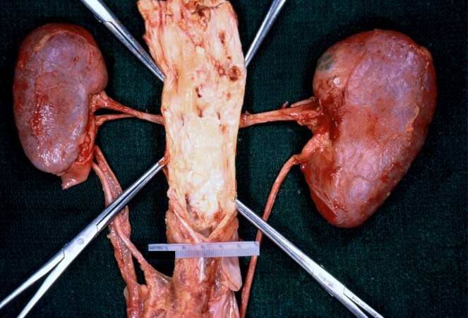
| 16:10, 19 August 2013 | IPLab2Atrophy7.jpg (file) |  |
50 KB | Peter Anderson | In this gross photograph of kidneys and the abdominal aorta, there is narrowing of the left renal artery at its ostium from the aorta. This atherosclerotic narrowing of the renal artery causes reduced blood pressure in the kidney whose artery is affect... | 1 |
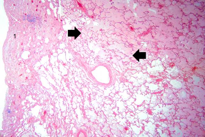
| 16:08, 19 August 2013 | IPLab4PulmonaryCongestion5.jpg (file) |  |
63 KB | Seung Park | This is a higher-power photomicrograph of lung. The edema fluid within the alveoli is visible at this higher magnification (arrows). The thickened pleura (1) is on the left. | 1 |
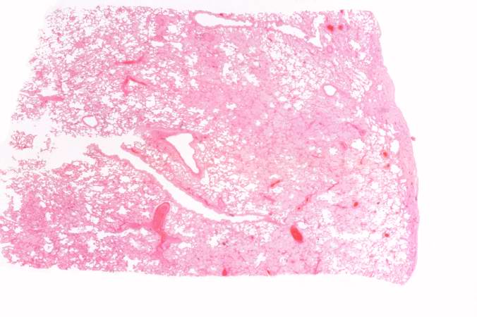
| 16:07, 19 August 2013 | IPLab4PulmonaryCongestion4.jpg (file) |  |
42 KB | Seung Park | This is a low-power photomicrograph of lung from this case. The lung section has a pale-red color indicating proteinaceous material within the lung. | 1 |
| 16:07, 19 August 2013 | IPLab4PulmonaryCongestion3.jpg (file) |  |
48 KB | Seung Park | This gross photograph demonstrates the frothy exudate that is being extruded from the lung tissue. | 1 |
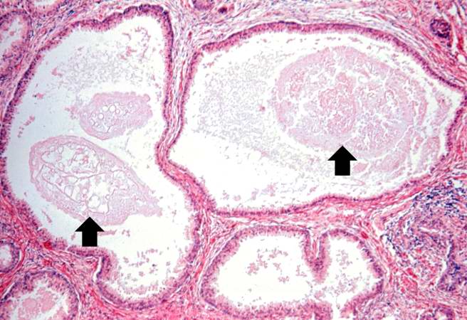
| 16:07, 19 August 2013 | IPLab2Atrophy6.jpg (file) |  |
52 KB | Peter Anderson | In this slide, the atrophy of the tubules is extending to include the Rete testes (arrow) as well. | 1 |
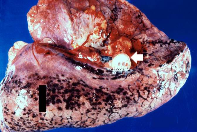
| 16:07, 19 August 2013 | IPLab4PulmonaryCongestion2.jpg (file) |  |
54 KB | Seung Park | This is a gross photograph of lung demonstrating acute pulmonary congestion and edema. A frothy exudate fills the bronchus (arrow). | 1 |
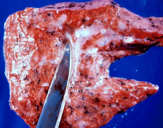
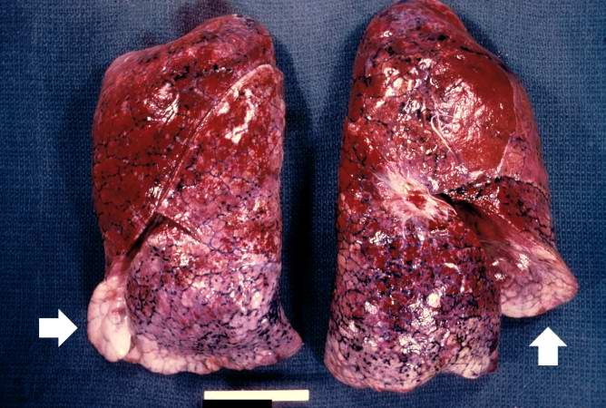
| 16:06, 19 August 2013 | IPLab4PulmonaryCongestion1.jpg (file) |  |
64 KB | Seung Park | This is a gross photograph of lungs that are distended and red. The reddish coloration of the tissue is due to congestion. Some normal pink lung tissue is seen at the edges of the lungs (arrows). | 1 |
| 16:04, 19 August 2013 | IPLab2Atrophy5.jpg (file) |  |
60 KB | Peter Anderson | A seminiferous tubule is shown on the left containing remnants of spermatocytes (1). There are no mature sperm present. On the right-hand portion of the slide are remnants of other spermatic tubules which have completely atrophied and lost all of their... | 1 |
| 16:03, 19 August 2013 | IPLab2Atrophy4.jpg (file) |  |
68 KB | Peter Anderson | This is a higher-power photomicrograph indicating loss of testicular parenchymal tissue. There are very few recognizable spermatic cells in this tissue. The cluster of cells in the upper right is a focus of interstitial or Leydig cells (arrow). These c... | 1 |
| 16:01, 19 August 2013 | IPLab2Atrophy3.jpg (file) |  |
65 KB | Peter Anderson | This is a higher-power photomicrograph of an atrophic testis. In this section there are seminiferous tubules with viable cells (1) although there are no visible spermatocytes. Other seminiferous tubules are completely acellular and have a pale pink hya... | 1 |
| 16:01, 19 August 2013 | IPLab2Atrophy2.jpg (file) |  |
53 KB | Peter Anderson | This is a low-power photomicrograph of an atrophic testis. Attached to the testis are several vessels (arrow) which are part of the epididymis and the vas deferens. | 1 |
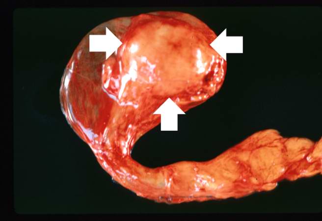
| 16:00, 19 August 2013 | IPLab2Atrophy1.jpg (file) |  |
23 KB | Peter Anderson | This is a gross photograph of an atrophied testis (arrows). | 1 |
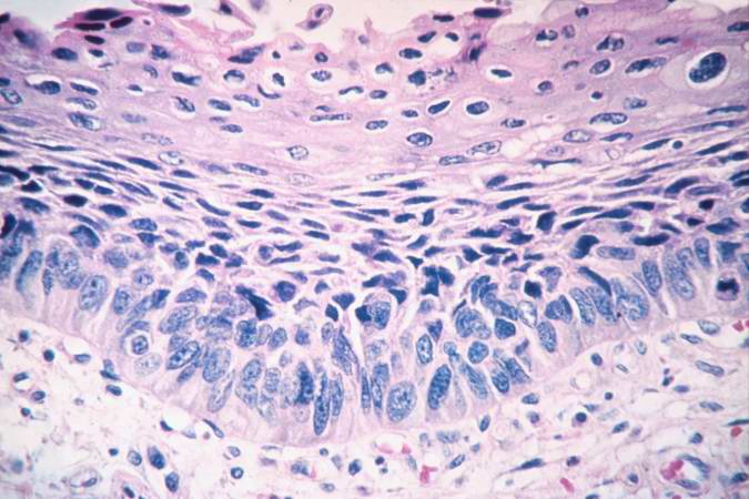
| 15:45, 19 August 2013 | IPLab2Metaplasia7.jpg (file) |  |
57 KB | Peter Anderson | This is a photomicrograph of the trachea from a smoker. Note that the columnar ciliated epithelium has been replaced by squamous epithelium. | 1 |
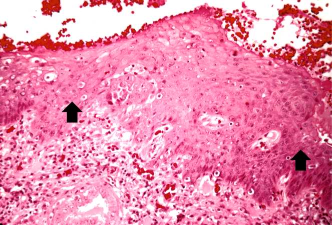
| 15:45, 19 August 2013 | IPLab2Metaplasia6.jpg (file) |  |
67 KB | Peter Anderson | A high-power photomicrograph of the squamous epithelium shows inflammatory cells in the subepithelial tissue and the formation of keratinized epithelium (arrows). | 1 |
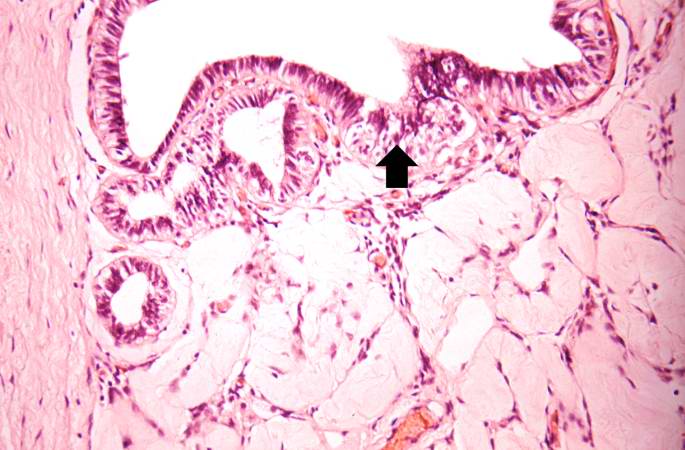
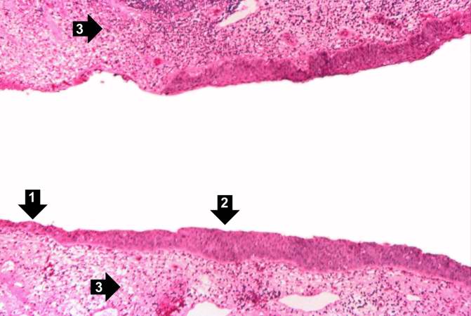
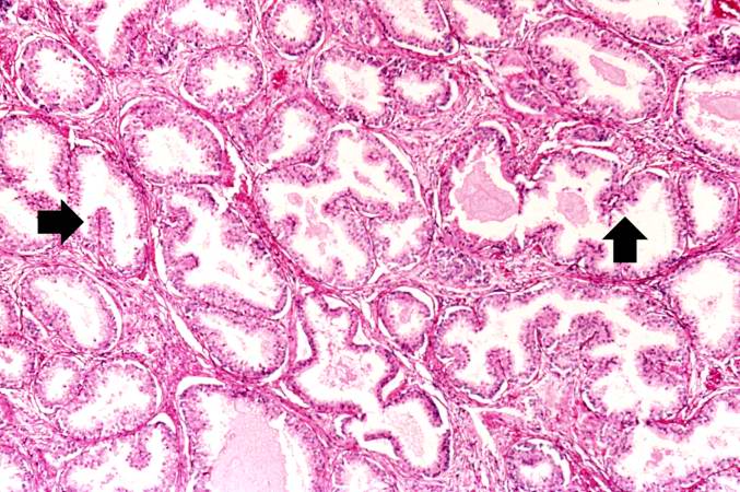
| 15:44, 19 August 2013 | IPLab2Metaplasia5.jpg (file) |  |
76 KB | Peter Anderson | In areas adjacent to the normal transitional epithelium, there are areas of epithelium (arrows) where the epithelial cells have the character of normal squamous epithelium as found in the dermis. However, squamous epithelium is not normal in the renal ... | 1 |
| 15:43, 19 August 2013 | IPLab2Metaplasia4.jpg (file) |  |
46 KB | Peter Anderson | This is a higher-power photomicrograph of the junction of normal epithelium (1) with hyperplastic transitional epithelium (2). | 1 |
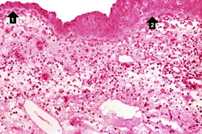
| 15:41, 19 August 2013 | IPLab2Metaplasia3.jpg (file) |  |
64 KB | Peter Anderson | A higher-power view shows the junction of normal epithelium (1) with hyperplastic transitional epithelium (2). Note the inflammatory cells in the subepithelial tissue. | 1 |
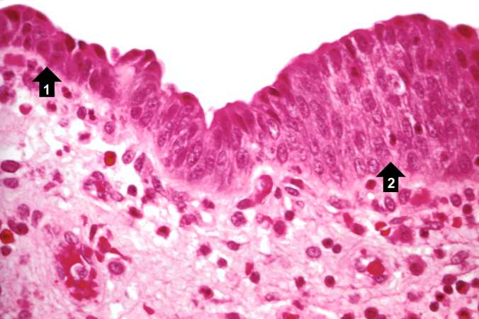
| 15:40, 19 August 2013 | IPLab2Metaplasia2.jpg (file) |  |
41 KB | Peter Anderson | his high-power photomicrograph demonstrates the transitional epithelium lining the renal calyx (1) and the junction (transition zone) to a thicker hyperplastic epithelium (2). Note the inflammatory cells and increased vascular response in the stromal t... | 1 |
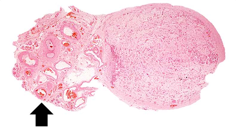
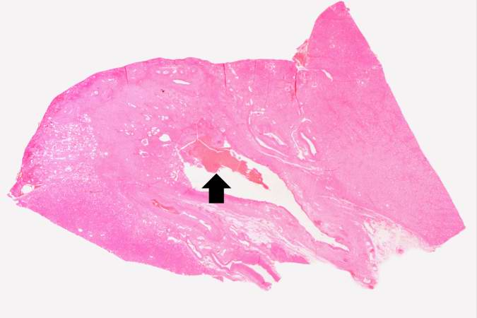
| 15:39, 19 August 2013 | IPLab2Metaplasia1.jpg (file) |  |
26 KB | Peter Anderson | This is a low-power photomicrograph showing the full cortical and medullary thickness of the kidney. Note that there is a dilated calyx containing some red blood cells in the center of the section (arrow). The cortex is markedly thin and has severe les... | 1 |
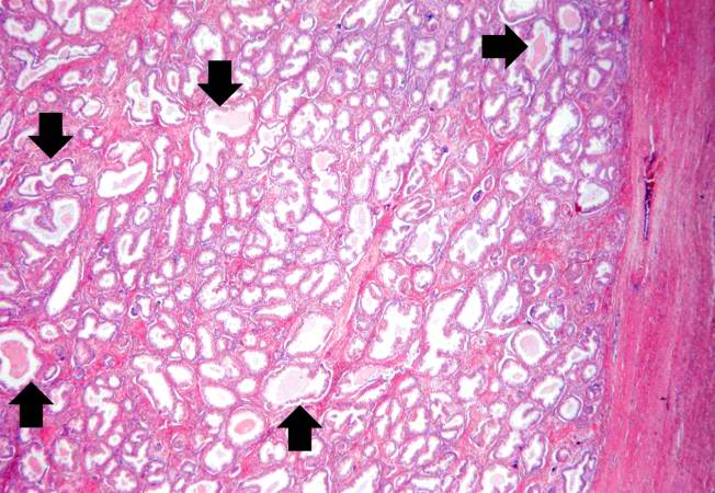
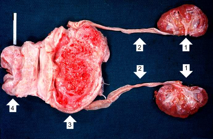
| 15:29, 19 August 2013 | IPLab2Hyperplasia9.jpg (file) |  |
37 KB | Peter Anderson | This kidney was removed from another autopsy patient who had prostatic hyperplasia resulting in marked urinary retention and back-flow of urine from the bladder into the ureters and renal pelvis. The increased pressure inside the renal pelvis resulted ... | 1 |
| 15:29, 19 August 2013 | IPLab2Hyperplasia8.jpg (file) |  |
53 KB | Peter Anderson | This is a higher-power photomicrograph of papillary folds of hyperplastic epithelium (arrows). | 1 |
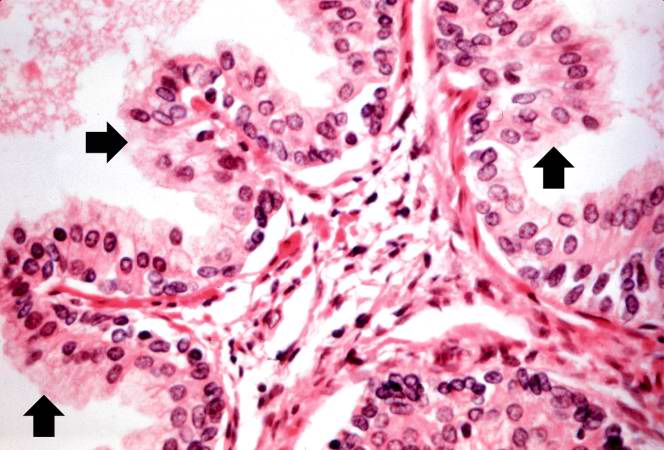
| 15:29, 19 August 2013 | IPLab2Hyperplasia7.jpg (file) |  |
74 KB | Peter Anderson | A higher-power view shows the papillary folds (arrows) produced by the hyperplastic epithelium projecting into the lumen of the gland. While these papillary folds project into the lumen of the gland, there is no extension through the glandular basement... | 1 |
| 15:28, 19 August 2013 | IPLab2Hyperplasia6.jpg (file) |  |
61 KB | Peter Anderson | Cystic dilatation of glands is present in this photomicrograph. Notice the accumulation of secretory material inside the glands (arrows) and compression (thinning) of the lining epithelium. | 1 |
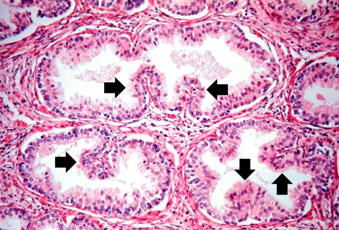
| 15:28, 19 August 2013 | IPLab2Hyperplasia5.jpg (file) |  |
86 KB | Peter Anderson | Note these glands, which exhibit hyperplasia of the glandular epithelium. The infolding of the glandular epithelial cells forms papillary projections (arrows) into the lumen of the gland. | 1 |
| 15:28, 19 August 2013 | IPLab2Hyperplasia4.jpg (file) |  |
69 KB | Peter Anderson | The dilated glands (arrows) make up the major portion of the prostate tissue and there is compression of the stroma. | 1 |
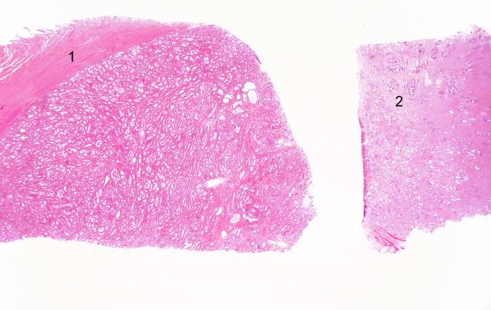
| 15:28, 19 August 2013 | IPLab2Hyperplasia3.jpg (file) |  |
48 KB | Peter Anderson | This is a low-power photomicrograph showing hyperplastic prostate on the left (1) and normal prostate on the right (2). At this power, dilated glands are visible in the section of hyperplastic prostate. | 1 |
| 15:27, 19 August 2013 | IPLab2Hyperplasia2.jpg (file) |  |
44 KB | Peter Anderson | This is a close-up of the prostate from this same patient. Note the nodularity of the tissue (1) and the enlargement of the gland. Enlargement of the prostate leads to compression of the urethra as it passes through (2) the gland. | 1 |
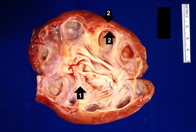
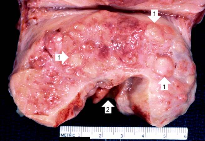
| 15:27, 19 August 2013 | IPLab2Hyperplasia1.jpg (file) |  |
58 KB | Peter Anderson | This photograph shows the autopsy specimen from this patient. Included are kidneys (1), ureters (2), bladder (3) (which has been opened), and enlarged prostate (4). Note that the bladder mucosa has multiple trabeculae and the bladder mucosa is hyperemi... | 1 |
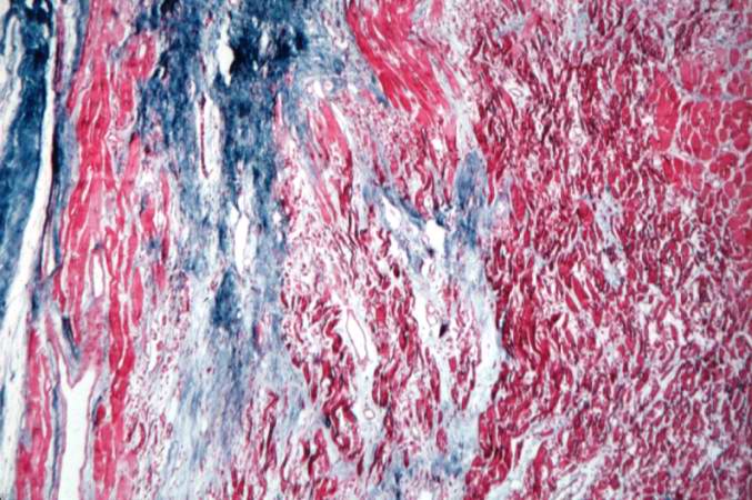
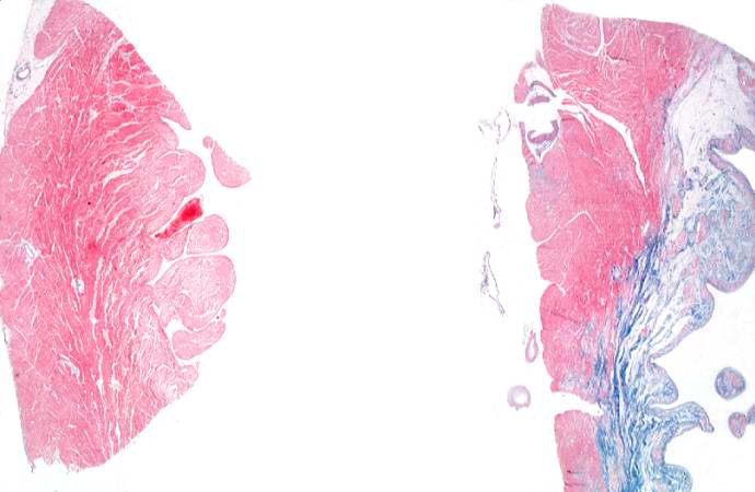
| 04:39, 19 August 2013 | IPLab3HealedMyocardialInfarction8.jpg (file) |  |
73 KB | Seung Park | This is a higher-power photomicrograph of a trichrome-stained section of heart containing an old healed MI. The scar tissue (mature fibrous connective tissue) is stained blue. | 1 |
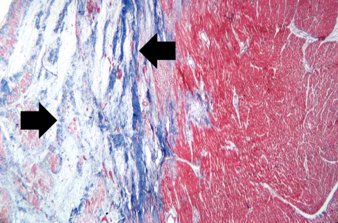
| 04:38, 19 August 2013 | IPLab3HealedMyocardialInfarction7.jpg (file) |  |
71 KB | Seung Park | This is a photomicrograph of a trichrome-stained section of heart containing an old healed myocardial infarction. The scar is composed of mature fibrous connective tissue (arrows). | 1 |
| 04:38, 19 August 2013 | IPLab3HealedMyocardialInfarction6.jpg (file) |  |
70 KB | Seung Park | This is a photomicrograph of a trichrome-stained section from a heart with an acute myocardial infarction. Note that there is little fibrous connective tissue. It is too early for scar formation to have taken place in this acute lesion. | 1 |
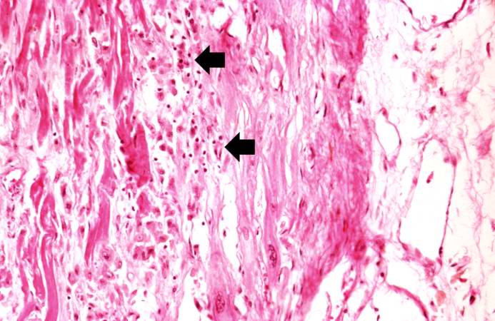
| 04:37, 19 August 2013 | IPLab3HealedMyocardialInfarction5.jpg (file) |  |
36 KB | Seung Park | This is a high-power photomicrograph of a different region of this healed MI. Note the chronic inflammatory reaction (arrows) in this region suggesting that there had been ischemic injury to this area within the last several weeks to months. | 1 |
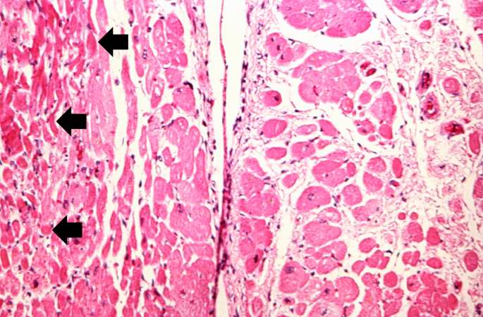
| 04:37, 19 August 2013 | IPLab3HealedMyocardialInfarction4.jpg (file) |  |
61 KB | Seung Park | This is another high-power photomicrograph of a healed myocardial infarction. Note the remaining normal myocytes (1), the fibrous connective tissue (2), and occasional hypereosinophilic myocytes indicating recent acute ischemic injury (arrow). | 1 |
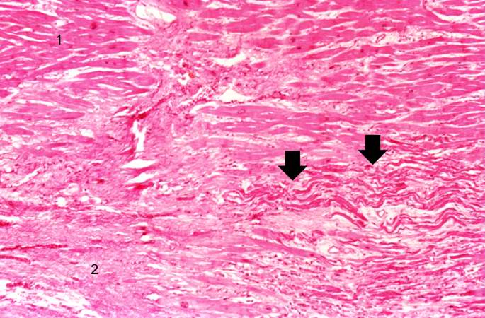
| 04:37, 19 August 2013 | IPLab3HealedMyocardialInfarction3.jpg (file) |  |
71 KB | Seung Park | This is a higher-power photomicrograph of a healed myocardial infarction with a fibrous scar. Remaining normal tissue is at the top (1) and the fibrous connective tissue scar is at the bottom (2). Note the presence of occasional hypereosinophilic myocy... | 1 |
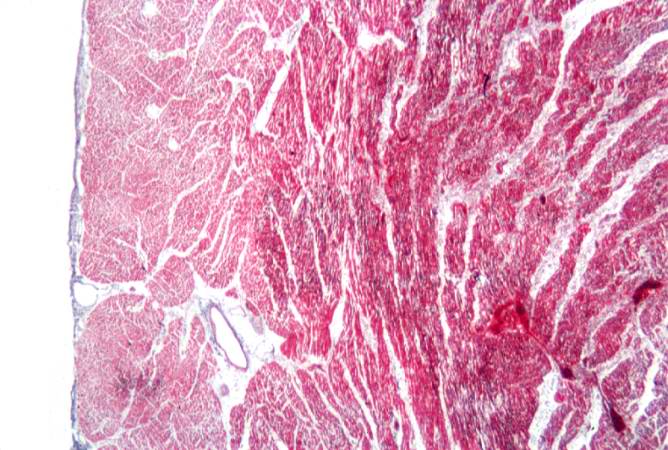
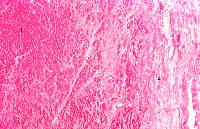
| 04:36, 19 August 2013 | IPLab3HealedMyocardialInfarction2.jpg (file) |  |
75 KB | Seung Park | This is a low-power photomicrograph of a healed myocardial infarction with a fibrous scar. Remaining normal tissue is on the left (1) and the fibrous connective tissue scar is on the right (2). | 1 |
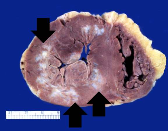
| 04:36, 19 August 2013 | IPLab3HealedMyocardialInfarction1.jpg (file) |  |
25 KB | Seung Park | This is a gross photograph of a heart with areas of old healed myocardial infarction (scars) outlined by arrows. | 1 |
| 04:30, 19 August 2013 | IPLab3AcuteMyocardialInfarction7.jpg (file) |  |
88 KB | Seung Park | This is a photomicrograph of the lines of Zahn. Pale areas (1) represent platelets with some fibrin and the darker lines (2) represent RBCs and leukocytes enmeshed in fibrin strands. | 1 |
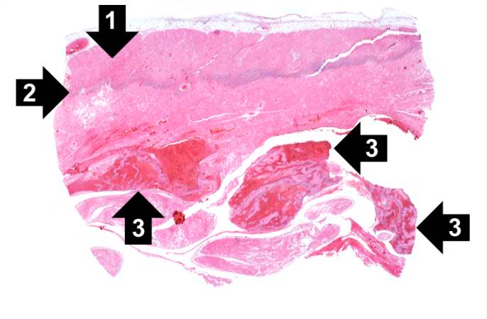
| 04:30, 19 August 2013 | IPLab3AcuteMyocardialInfarction6.jpg (file) |  |
79 KB | Seung Park | This is a low-power photomicrograph of a mural thrombus (1) adherent to the endocardial surface (arrows). | 1 |
| 04:30, 19 August 2013 | IPLab3AcuteMyocardialInfarction5.jpg (file) |  |
68 KB | Seung Park | This is a high-power photomicrograph of another area of this section. There are several hypereosinophilic cells within this section (arrows). | 1 |
| 04:30, 19 August 2013 | IPLab3AcuteMyocardialInfarction4.jpg (file) |  |
79 KB | Seung Park | This is a higher-power photomicrograph of the edge of the infarct. The accumulation of inflammatory cells is on the left (1) and the infarcted tissue is on the right (2). Note that intact cells can be seen in the infarct but there are no nuclei. | 1 |
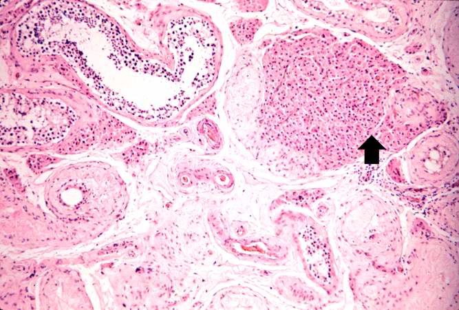
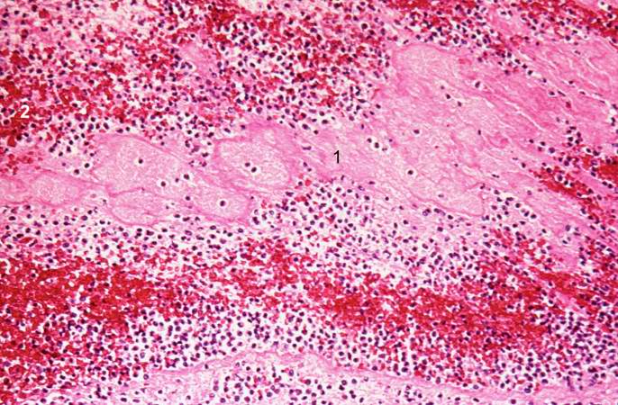
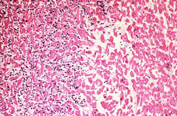
| 04:29, 19 August 2013 | IPLab3AcuteMyocardialInfarction3.jpg (file) |  |
83 KB | Seung Park | This is a photomicrograph of the edge of the infarct with normal tissue on the left (1). The accumulation of inflammatory cells (2) is at the edge of the infarcted tissue (3). | 1 |
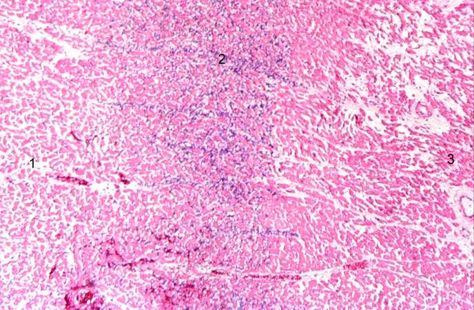
| 04:29, 19 August 2013 | IPLab3AcuteMyocardialInfarction2.jpg (file) |  |
60 KB | Seung Park | This is a higher-power photomicrograph which shows more clearly the viable tissue along the epicardium (1), the blue line of inflammatory cells (2), and the infarcted myocardium (3). | 1 |
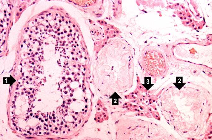
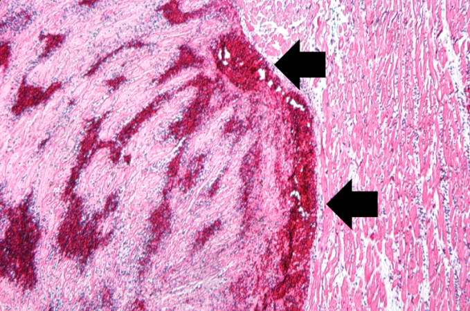
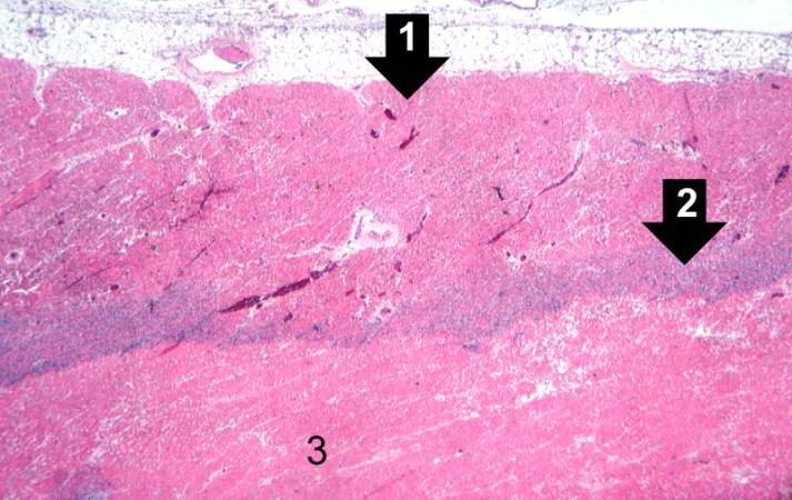
| 04:29, 19 August 2013 | IPLab3AcuteMyocardialInfarction1.jpg (file) |  |
36 KB | Seung Park | This is a low-power photomicrograph of infarcted heart. There is a layer of surviving myocardial tissue (1) along the epicardium and then a blue line (2) which represents the accumulation of inflammatory cells at the border of the infarct. There is thr... | 1 |
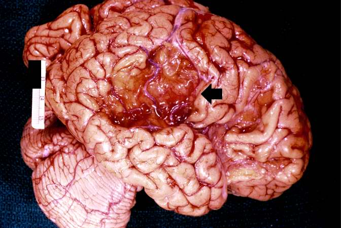
| 04:24, 19 August 2013 | IPLab3BrainInfarction11.jpg (file) |  |
56 KB | Seung Park | This is a closer view of the brain demonstrating an old healed infarct with the meninges containing blood vessels (arrow) overlying the infarcted region. | 1 |
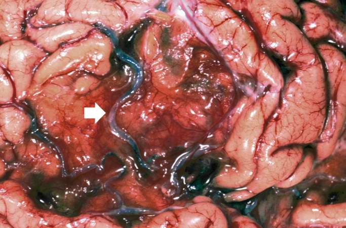
| 04:24, 19 August 2013 | IPLab3BrainInfarction10.jpg (file) |  |
56 KB | Seung Park | This is a gross photograph of a brain from another patient with an old healed infarct. Note the meninges overlying the infarcted region (arrow). | 1 |
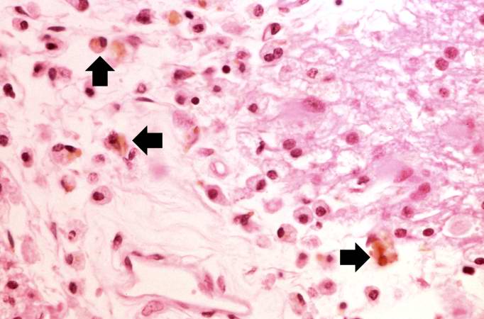
| 04:23, 19 August 2013 | IPLab3BrainInfarction9.jpg (file) |  |
43 KB | Seung Park | This is a photomicrograph of the edge of the infarct. Note the gitter cells, gemistocytic astrocytes, and some hemosiderin-laden macrophages (arrows). | 1 |
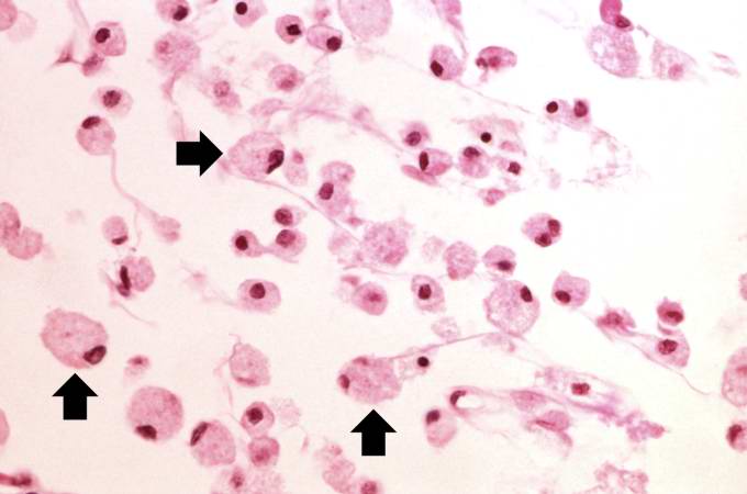
| 04:23, 19 August 2013 | IPLab3BrainInfarction8.jpg (file) |  |
28 KB | Seung Park | This is a high-power photomicrograph of gitter cells (arrows). | 1 |