File:IPLab4MuralThrombus6.jpg
Revision as of 16:29, 19 August 2013 by Seung Park (talk | contribs) (This is a higher-power photomicrograph of the thrombus. Note the pale regions which contain primarily platelets (degranulated platelets) with some fibrin (1), and the red areas which contain RBCs, some leukocytes, and fibrin(2).)
No higher resolution available.
IPLab4MuralThrombus6.jpg (671 × 450 pixels, file size: 82 KB, MIME type: image/jpeg)
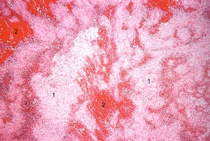
This is a higher-power photomicrograph of the thrombus. Note the pale regions which contain primarily platelets (degranulated platelets) with some fibrin (1), and the red areas which contain RBCs, some leukocytes, and fibrin(2).
A thrombus is a solid mass resulting from the aggregation of blood constituents within the vascular system.
File history
Click on a date/time to view the file as it appeared at that time.
| Date/Time | Thumbnail | Dimensions | User | Comment | |
|---|---|---|---|---|---|
| current | 16:29, 19 August 2013 | | 671 × 450 (82 KB) | Seung Park (talk | contribs) | This is a higher-power photomicrograph of the thrombus. Note the pale regions which contain primarily platelets (degranulated platelets) with some fibrin (1), and the red areas which contain RBCs, some leukocytes, and fibrin(2). |
- You cannot overwrite this file.
File usage
The following page links to this file: