File:IPLab7Carcinoid10.jpg
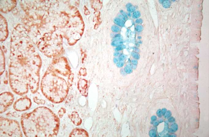
Revision as of 02:08, 21 August 2013 by Seung Park (talk | contribs) (This is a higher-power view of the previous section stained with a silver stain to delineate carcinoid tumor cells (brown) and a mucin stain (blue) to stain the glands.)
No higher resolution available.
IPLab7Carcinoid10.jpg (682 × 450 pixels, file size: 45 KB, MIME type: image/jpeg)
This is a higher-power view of the previous section stained with a silver stain to delineate carcinoid tumor cells (brown) and a mucin stain (blue) to stain the glands.
File history
Click on a date/time to view the file as it appeared at that time.
| Date/Time | Thumbnail | Dimensions | User | Comment | |
|---|---|---|---|---|---|
| current | 02:08, 21 August 2013 | | 682 × 450 (45 KB) | Seung Park (talk | contribs) | This is a higher-power view of the previous section stained with a silver stain to delineate carcinoid tumor cells (brown) and a mucin stain (blue) to stain the glands. |
- You cannot overwrite this file.
File usage
The following page links to this file: