File:IPLab8HSVGlossitis4.jpg
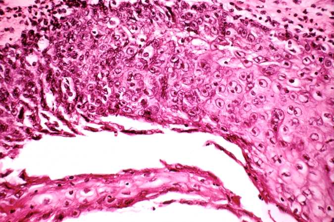
Revision as of 02:29, 21 August 2013 by Seung Park (talk | contribs) (This is a higher-power photomicrograph of epithelium at the edge of the ulcer. Amphophilic intranuclear inclusion bodies can be seen in almost all of the epithelial cells in this section.)
No higher resolution available.
IPLab8HSVGlossitis4.jpg (675 × 450 pixels, file size: 72 KB, MIME type: image/jpeg)
This is a higher-power photomicrograph of epithelium at the edge of the ulcer. Amphophilic intranuclear inclusion bodies can be seen in almost all of the epithelial cells in this section.
File history
Click on a date/time to view the file as it appeared at that time.
| Date/Time | Thumbnail | Dimensions | User | Comment | |
|---|---|---|---|---|---|
| current | 02:29, 21 August 2013 | | 675 × 450 (72 KB) | Seung Park (talk | contribs) | This is a higher-power photomicrograph of epithelium at the edge of the ulcer. Amphophilic intranuclear inclusion bodies can be seen in almost all of the epithelial cells in this section. |
- You cannot overwrite this file.
File usage
The following page links to this file: