File:IPLab8HSVEncephalitis6.jpg
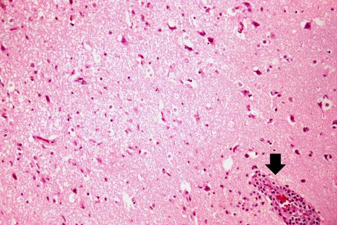
Revision as of 02:35, 21 August 2013 by Seung Park (talk | contribs) (This is another high-power photomicrograph showing a blood vessel with perivascular hemorrhage and mild perivascular lymphocytic cuffing (arrow). In addition, there are numerous red shrunken neurons and glia with pyknotic nuclei throughout this section.)
No higher resolution available.
IPLab8HSVEncephalitis6.jpg (673 × 450 pixels, file size: 67 KB, MIME type: image/jpeg)
This is another high-power photomicrograph showing a blood vessel with perivascular hemorrhage and mild perivascular lymphocytic cuffing (arrow). In addition, there are numerous red shrunken neurons and glia with pyknotic nuclei throughout this section.
File history
Click on a date/time to view the file as it appeared at that time.
| Date/Time | Thumbnail | Dimensions | User | Comment | |
|---|---|---|---|---|---|
| current | 02:35, 21 August 2013 | | 673 × 450 (67 KB) | Seung Park (talk | contribs) | This is another high-power photomicrograph showing a blood vessel with perivascular hemorrhage and mild perivascular lymphocytic cuffing (arrow). In addition, there are numerous red shrunken neurons and glia with pyknotic nuclei throughout this section. |
- You cannot overwrite this file.
File usage
The following page links to this file: