File:IPLab9RMSF2.jpg
Revision as of 03:42, 21 August 2013 by Seung Park (talk | contribs) (This is a higher-power photomicrograph demonstrating areas of hemorrhage immediately underneath the epidermis. Also note the cellularity and thrombosis of the small vessels in the dermis (arrow).)
No higher resolution available.
IPLab9RMSF2.jpg (666 × 450 pixels, file size: 51 KB, MIME type: image/jpeg)
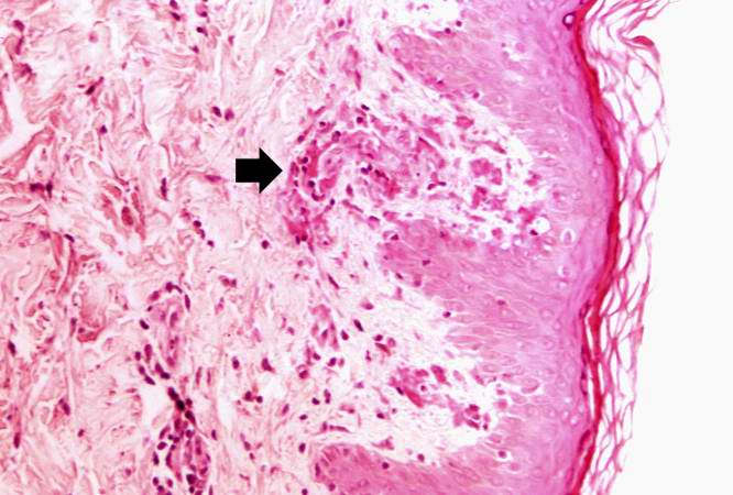
This is a higher-power photomicrograph demonstrating areas of hemorrhage immediately underneath the epidermis. Also note the cellularity and thrombosis of the small vessels in the dermis (arrow).
File history
Click on a date/time to view the file as it appeared at that time.
| Date/Time | Thumbnail | Dimensions | User | Comment | |
|---|---|---|---|---|---|
| current | 03:42, 21 August 2013 | | 666 × 450 (51 KB) | Seung Park (talk | contribs) | This is a higher-power photomicrograph demonstrating areas of hemorrhage immediately underneath the epidermis. Also note the cellularity and thrombosis of the small vessels in the dermis (arrow). |
- You cannot overwrite this file.
File usage
The following page links to this file: