File list
This special page shows all uploaded files.
| Date | Name | Thumbnail | Size | User | Description | Versions |
|---|---|---|---|---|---|---|
| 05:13, 21 August 2013 | IPLab12Alcoholic6.jpg (file) |  |
42 KB | Seung Park | In this high-power photomicrograph of trichrome-stained liver, the bands of fibrous tissue surround the hepatocyte nodules. There is some degeneration and dropout of hepatocytes in this nodule. Also note the increased numbers of bile ducts in the triad... | 1 |
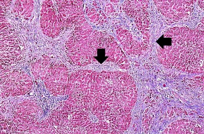
| 05:13, 21 August 2013 | IPLab12Alcoholic4.jpg (file) |  |
130 KB | Seung Park | In this is medium-power photomicrograph of trichrome stained liver the bands of fibrous tissue are seen to form "bridges" between triad areas (arrows); this is called "bridging fibrosis." Also note the fibrous tissue (arrows) and how the hepatocytes ar... | 1 |
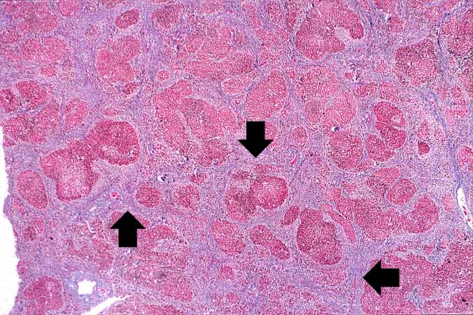
| 05:13, 21 August 2013 | IPLab12Alcoholic3.jpg (file) |  |
102 KB | Seung Park | This is a low-power photomicrograph of this liver stained with a trichrome stain to highlight the fibrous tissue (arrows). Also note the nodular pattern. | 1 |
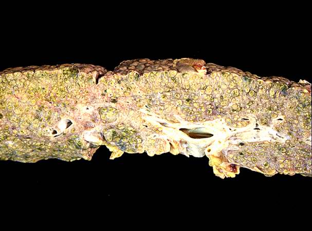
| 05:13, 21 August 2013 | IPLab12Alcoholic2.jpg (file) |  |
65 KB | Seung Park | This is a closer view of the liver from this patient. Again note the nodular pattern and the areas of greenish discoloration. These green nodules are actually the viable hepatocytes that are stained green because of bile stasis. The pale areas are the ... | 1 |
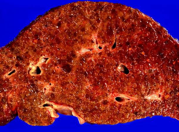
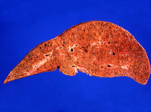
| 05:13, 21 August 2013 | IPLab12Alcoholic1.jpg (file) |  |
40 KB | Seung Park | This is a gross photograph of the liver from this patient. Note the nodular pattern and the areas of greenish discoloration as well as pale tan areas. | 1 |
| 05:08, 21 August 2013 | IPLab11Chagas6.jpg (file) |  |
34 KB | Seung Park | This is a higher-power photomicrograph of an H & E stained heart biopsy from this patient. Note the T. cruzi amastigotes (arrows) within this longitudinal section of a myocyte. | 1 |
| 05:08, 21 August 2013 | IPLab11Chagas5.jpg (file) |  |
48 KB | Seung Park | This is a higher-power photomicrograph of an H & E stained heart biopsy from this patient. At this magnification the organisms within a myocyte (arrows) and the adjacent inflammatory response are more clearly seen. The individual organisms within the m... | 1 |
| 05:07, 21 August 2013 | IPLab11Chagas4.jpg (file) |  |
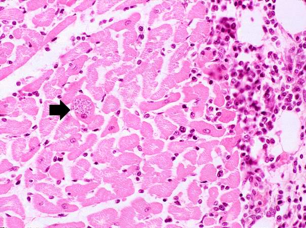
63 KB | Seung Park | This is a higher-power photomicrograph of an H & E stained heart biopsy from this patient. Again, note the organisms within a myocyte (arrow) and the inflammatory response. | 1 |
| 05:07, 21 August 2013 | IPLab11Chagas3.jpg (file) |  |
64 KB | Seung Park | This is a low-power photomicrograph of an H & E stained section from the heart biopsy of this patient. Note the organisms within a myocyte (arrow) and the adjacent inflammatory response. | 1 |
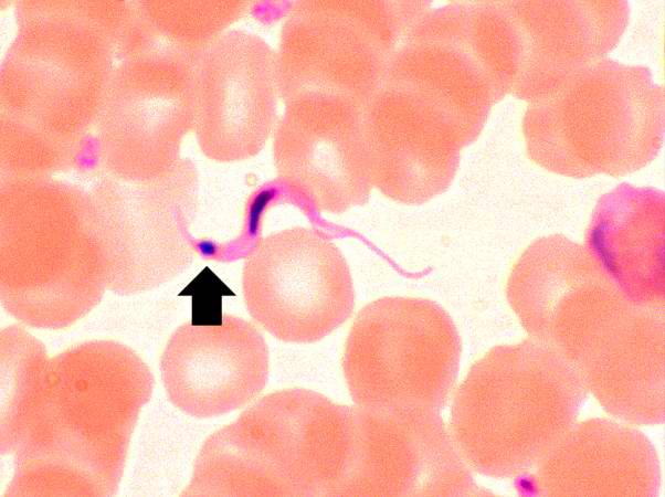
| 05:07, 21 August 2013 | IPLab11Chagas2.jpg (file) |  |
34 KB | Seung Park | This peripheral blood smear from the patient shows a higher power view of a Trypanosoma cruzi trypomastigote. Note the prominent kinetoplast (arrow). | 1 |
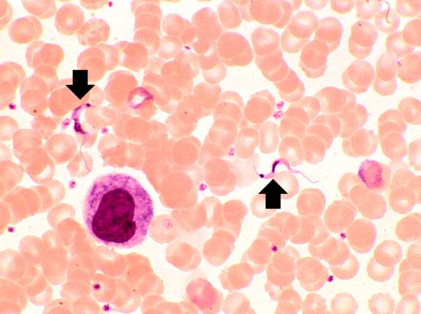
| 05:07, 21 August 2013 | IPLab11Chagas1.jpg (file) |  |
37 KB | Seung Park | This peripheral blood smear from the patient shows two trypomastigotes of Trypanosoma cruzi. | 1 |
| 05:05, 21 August 2013 | IPLab11Schistosomiasis3.jpg (file) |  |
79 KB | Seung Park | This is a photomicrograph of the liver from another patient with Schistosoma mansoni. These eggs have elicited a robust inflammatory response, including a multinucleated giant cell. These lesions go on to heal by fibrosis resulting in cirrhosis and the... | 1 |
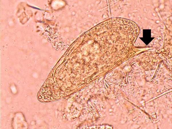
| 05:05, 21 August 2013 | IPLab11Schistosomiasis2.jpg (file) |  |
41 KB | Seung Park | This is high-power photomicrograph of the patient's fecal specimen containing another Schistosoma mansoni egg. | 1 |
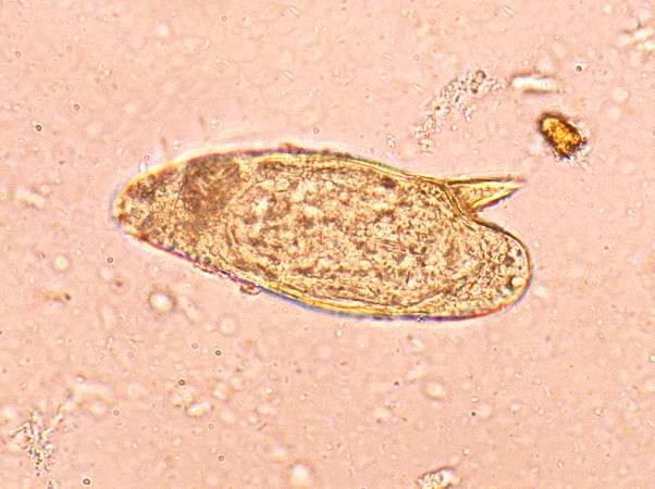
| 05:05, 21 August 2013 | IPLab11Schistosomiasis1.jpg (file) |  |
53 KB | Seung Park | This is a high-power photomicrograph of the patient's fecal specimen containing the characteristic egg of Schistosoma mansoni. Note the spine on the egg (arrow). | 1 |
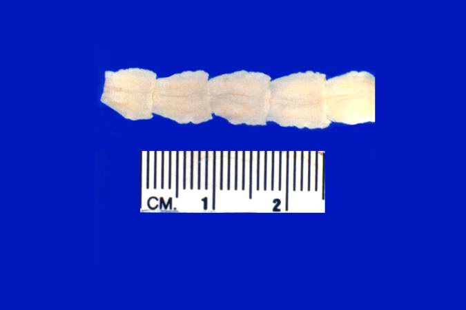
| 05:04, 21 August 2013 | IPLab11Cysticercosis4.jpg (file) |  |
13 KB | Seung Park | This is a photograph of the body of an adult tapeworm. Note the body segments. | 1 |
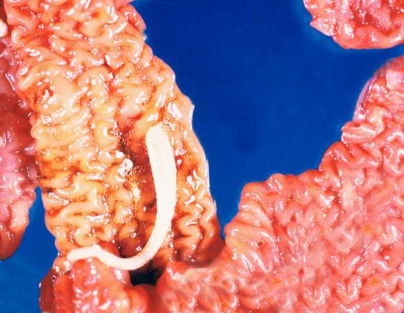
| 05:04, 21 August 2013 | IPLab11Cysticercosis3.jpg (file) |  |
46 KB | Seung Park | This photograph of an autopsy specimen from another patient shows an adult tapeworm in the intestine. Note that the worm attaches to the luminal surface of the intestine via the scolex. | 1 |
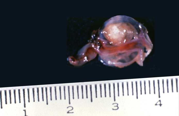
| 05:03, 21 August 2013 | IPLab11Cysticercosis2.jpg (file) |  |
20 KB | Seung Park | This is a photograph of the cyst that was surgically removed. The cyst is filled with a clear fluid and contains a scolex. | 1 |
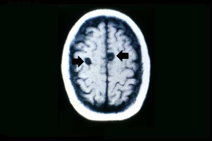
| 05:03, 21 August 2013 | IPLab11Cysticercosis1.jpg (file) |  |
17 KB | Seung Park | This is a head CT showing the two cysts (arrows). | 1 |
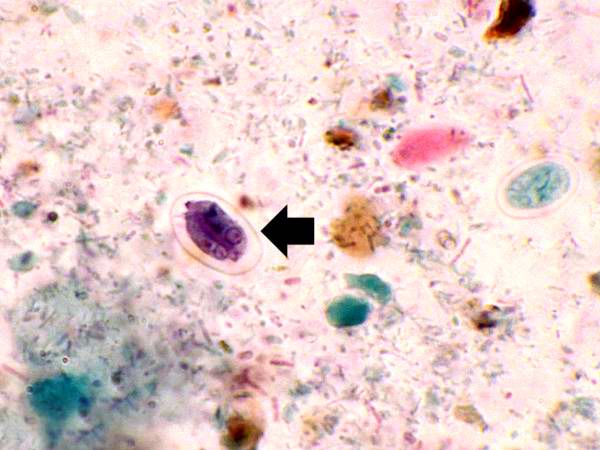
| 05:02, 21 August 2013 | IPLab11Ascariasis7.jpg (file) |  |
40 KB | Seung Park | This high-power photomicrograph of the fecal specimen from this patient shows a Giardia lamblia cyst (arrow). | 1 |
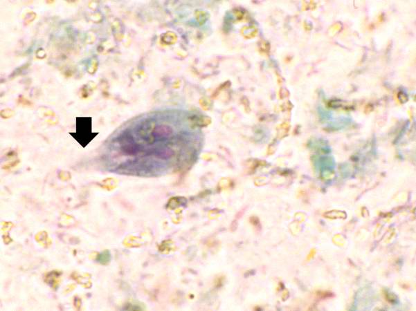
| 05:01, 21 August 2013 | IPLab11Ascariasis6.jpg (file) |  |
29 KB | Seung Park | This high-power photomicrograph of the fecal specimen from this patient shows a Giardia lamblia trophozoite. Note the two nuclei and the tapered end (that goes out of the plane of focus) containing flagella (arrow). | 1 |
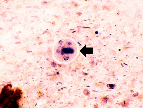
| 05:01, 21 August 2013 | IPLab11Ascariasis5.jpg (file) |  |
41 KB | Seung Park | This high-power photomicrograph of the fecal specimen from this same patient shows an Entamoeba histolytica cyst (arrow). | 1 |
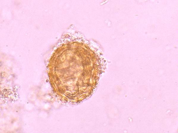
| 05:01, 21 August 2013 | IPLab11Ascariasis4.jpg (file) |  |
22 KB | Seung Park | This is a higher-power photomicrograph of another ascarid egg. | 1 |
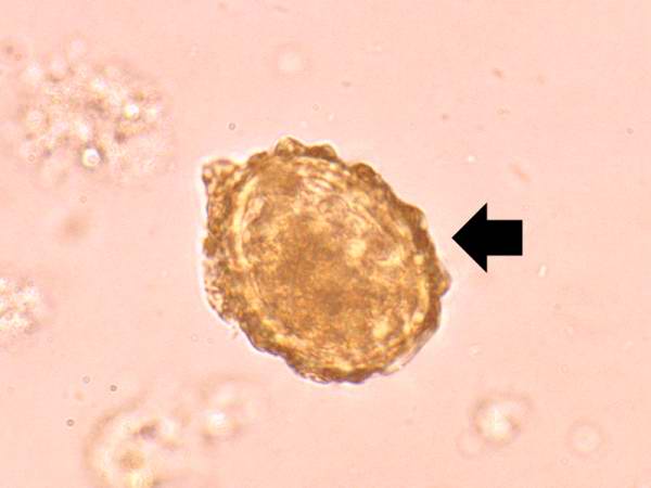
| 05:01, 21 August 2013 | IPLab11Ascariasis3.jpg (file) |  |
19 KB | Seung Park | This is a high-power photomicrograph of a fecal specimen from this patient showing an ascarid egg (arrow). | 1 |
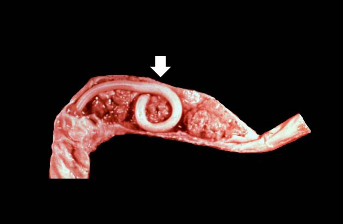
| 05:01, 21 August 2013 | IPLab11Ascariasis2.jpg (file) |  |
16 KB | Seung Park | This is a photograph of an autopsy specimen from another case of ascariasis. The adult ascarid (arrow) can be seen in the section of small bowel from this patient. | 1 |
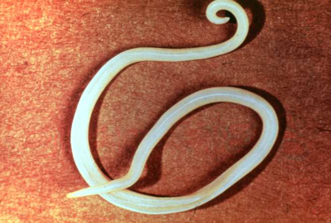
| 05:00, 21 August 2013 | IPLab11Ascariasis1.jpg (file) |  |
45 KB | Seung Park | This is a photograph of an adult ascarid like the ones removed from the small bowel of this patient. | 1 |
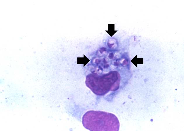
| 04:58, 21 August 2013 | IPLab11Leishmaniasis6.jpg (file) |  |
19 KB | Seung Park | This is a high-power photomicrograph of a touch prep made from the skin lesion at the time of biopsy. A single macrophage can be seen with intracytoplasmic leishmania organisms (arrows). | 1 |
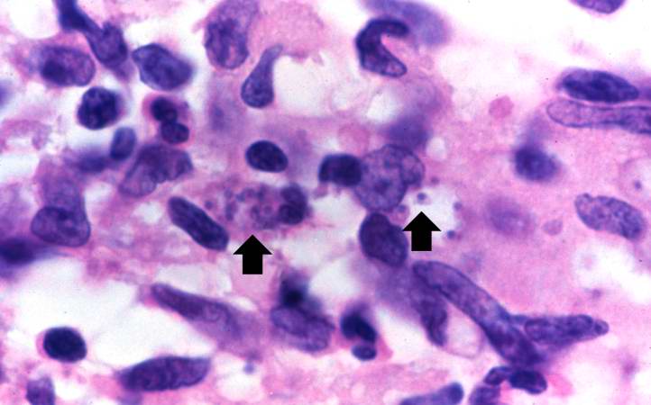
| 04:58, 21 August 2013 | IPLab11Leishmaniasis5.jpg (file) |  |
38 KB | Seung Park | This is a high-power photomicrograph of an inflammatory cell containing cytoplasmic organisms (arrows). | 1 |
| 04:58, 21 August 2013 | IPLab11Leishmaniasis4.jpg (file) |  |
70 KB | Seung Park | This high-power photomicrograph of the biopsy specimen shows more clearly the heavily infiltrate of inflammatory cells. Note the small blue structures inside the inflammatory cells (arrows). | 1 |
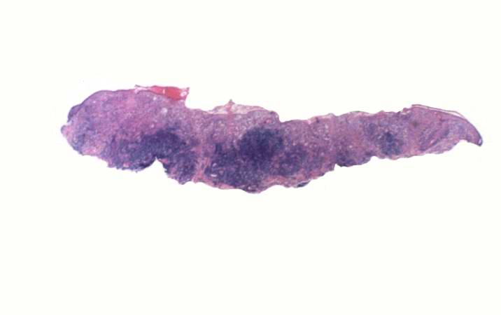
| 04:58, 21 August 2013 | IPLab11Leishmaniasis3.jpg (file) |  |
58 KB | Seung Park | This is a higher-power photomicrograph of this biopsy. The ulcerated surface is seen on the top of the section. Again, note that the specimen is heavily infiltrated with inflammatory cells. | 1 |
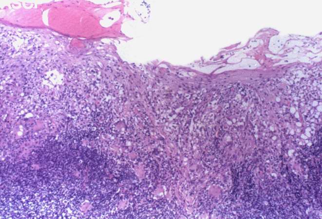
| 04:58, 21 August 2013 | IPLab11Leishmaniasis2.jpg (file) |  |
13 KB | Seung Park | This is a low-power photomicrograph of the biopsy taken from this skin lesion. The ulcerated surface is at the top. Note that the specimen is heavily infiltrated with inflammatory cells. | 1 |
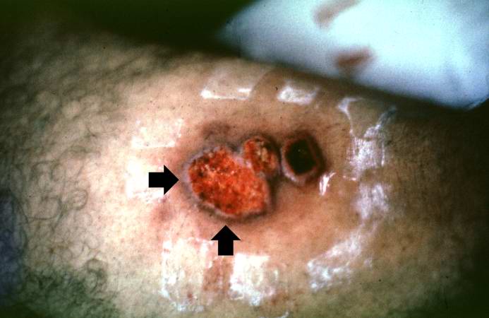
| 04:57, 21 August 2013 | IPLab11Leishmaniasis1.jpg (file) |  |
34 KB | Seung Park | In this photograph of the skin lesion seen in this patient, note the raised edges (arrows) and the ulcerated center. | 1 |
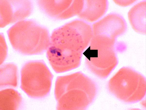
| 04:56, 21 August 2013 | IPLab11Malaria7.jpg (file) |  |
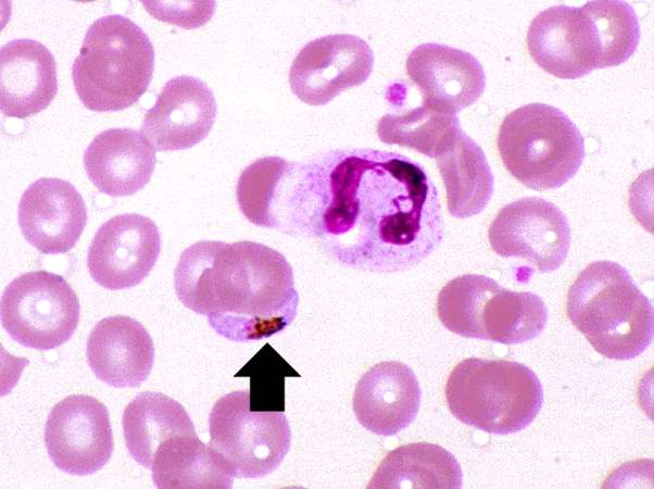
24 KB | Seung Park | In this peripheral smear from a different patient who was infected with P. vivax, the cytoplasm of the infected RBC has a stippled appearance (Schüffner's dots) (arrow). The RBC is also slightly enlarged. | 1 |
| 04:55, 21 August 2013 | IPLab11Malaria6.jpg (file) |  |
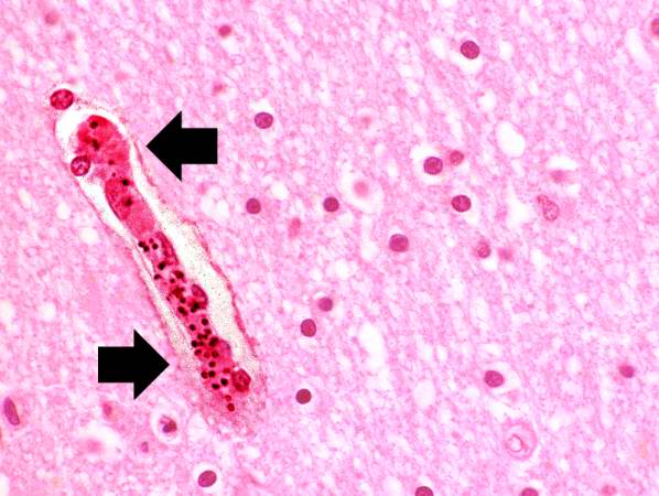
47 KB | Seung Park | This high photomicrograph was taken from another patient who died of malignant cerebral malaria caused by P. falciparum. In this photomicrograph, a small artery (arrow) can be seen that is full of parasitized RBCs. These RBCs tend to clog small blood v... | 1 |
| 04:55, 21 August 2013 | IPLab11Malaria5.jpg (file) |  |
37 KB | Seung Park | There is another example of a P. falciparum gametocyte (arrow) in this thin smear. There is a neutrophil in this field as well. | 1 |
| 04:55, 21 August 2013 | IPLab11Malaria4.jpg (file) |  |
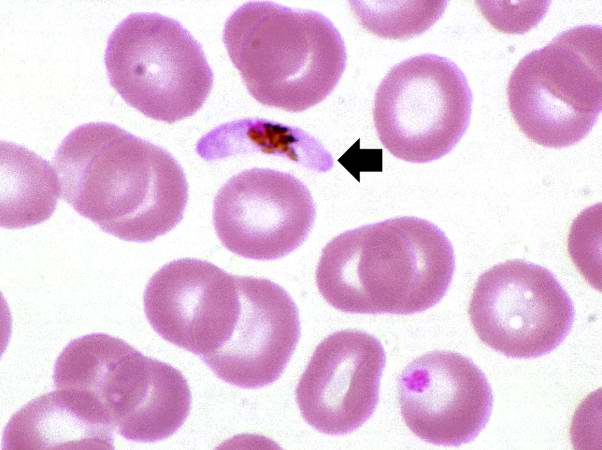
30 KB | Seung Park | In this high power photomicrograph of a thin smear of blood from this patient there is one P. falciparum gametocyte (arrow). These gametocytes have a characteristic "banana" shape. | 1 |
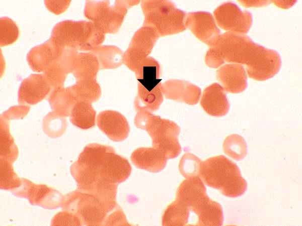
| 04:55, 21 August 2013 | IPLab11Malaria3.jpg (file) |  |
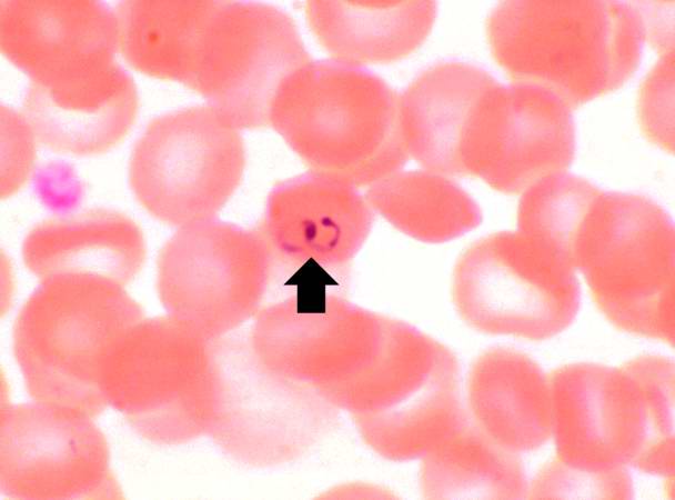
22 KB | Seung Park | This is yet another high power photomicrograph of a thin smear of blood from this patient. There is one RBC that contains two ring stage trophozoites (arrow). This is characteristic of, but not diagnostic for, P. falciparum. | 1 |
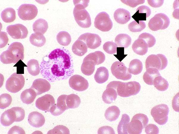
| 04:54, 21 August 2013 | IPLab11Malaria2.jpg (file) |  |
36 KB | Seung Park | This is another high power photomicrograph of a thin smear of blood from this patient. There is a single eosinophil in this smear along with several RBCs containing ring stage trophozoites (arrows). | 1 |
| 04:54, 21 August 2013 | IPLab11Malaria1.jpg (file) |  |
24 KB | Seung Park | This is a high power photomicrograph of a thin smear of blood from this patient. Note that one of the RBCs has a ring stage trophozoite (arrow). | 1 |
| 04:20, 21 August 2013 | IPLab10Mucor8.jpg (file) |  |
50 KB | Seung Park | This is another high-power photomicrograph of the thrombus stained to outline the Mucor organisms (arrows). | 1 |
| 04:20, 21 August 2013 | IPLab10Mucor7.jpg (file) |  |
51 KB | Seung Park | This is an even higher-power photomicrograph of the thrombus stained to outline the Mucor organisms (arrows). | 1 |
| 04:20, 21 August 2013 | IPLab10Mucor6.jpg (file) |  |
76 KB | Seung Park | This medium-power photomicrograph shows the thrombus stained to outline the Mucor organisms (arrows). Note again the ribbon-like morphology and the wide-angle branching. | 1 |
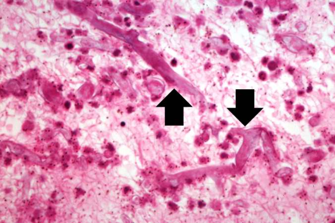
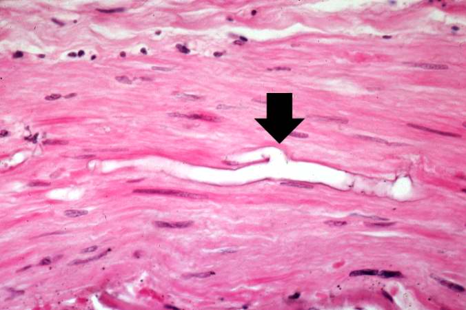
| 04:20, 21 August 2013 | IPLab10Mucor5.jpg (file) |  |
61 KB | Seung Park | This is another high-power photomicrograph of the wall of the artery and the thrombus. Within the thrombus there are multiple variably-shaped clear areas that represent longitudinal sections and cross sections of the Mucor organisms (arrows). | 1 |
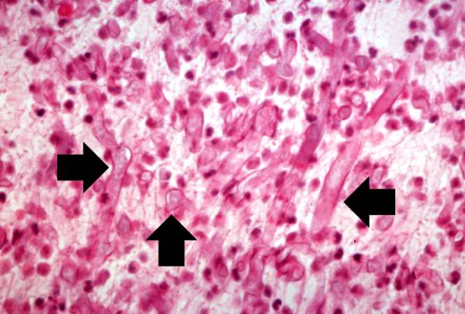
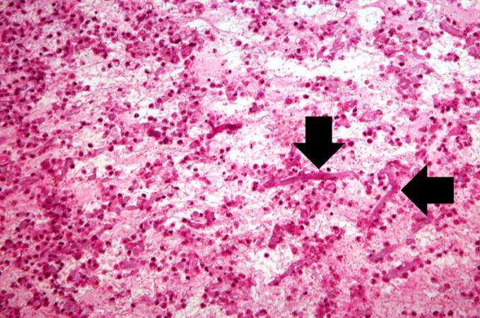
| 04:19, 21 August 2013 | IPLab10Mucor4.jpg (file) |  |
40 KB | Seung Park | This is a higher-power photomicrograph of just the wall of the carotid artery. Note the ribbon-like clear structure with roughly parallel walls (non-septate hyphae) and right-angle branching (arrow). This is the Mucor organism. | 1 |
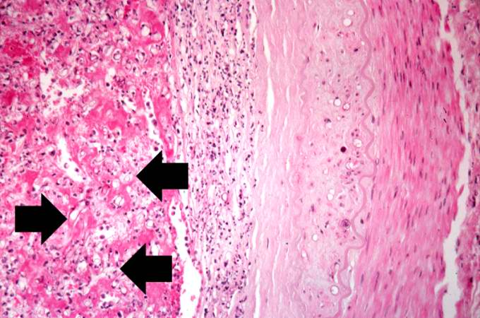
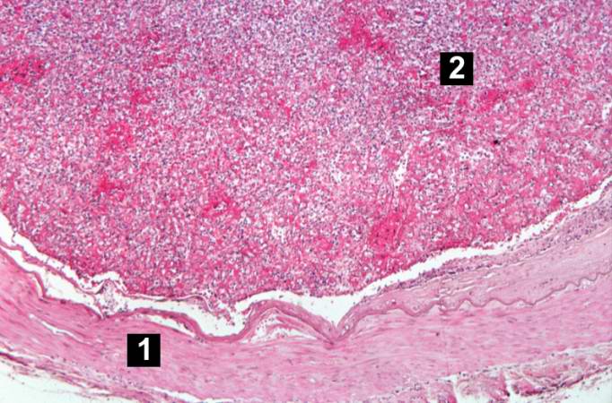
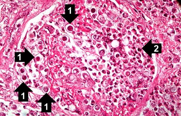
| 04:19, 21 August 2013 | IPLab10Mucor3.jpg (file) |  |
70 KB | Seung Park | This is an even higher-power photomicrograph of the wall of the carotid artery (1) and the thrombus (2). Within the wall of the artery and in the thrombus there are multiple variably shaped clear areas (3). At this magnification and with this stain, it... | 1 |
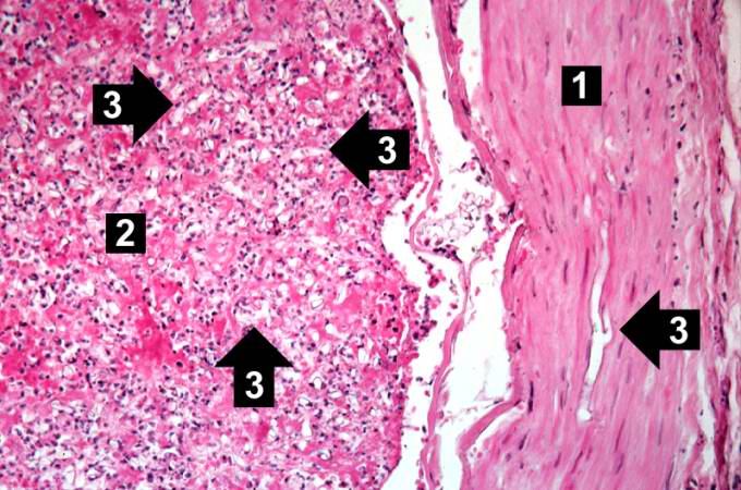
| 04:19, 21 August 2013 | IPLab10Mucor2.jpg (file) |  |
70 KB | Seung Park | This is a higher-power photomicrograph of the wall of the carotid artery (1) and the thrombus (2). | 1 |
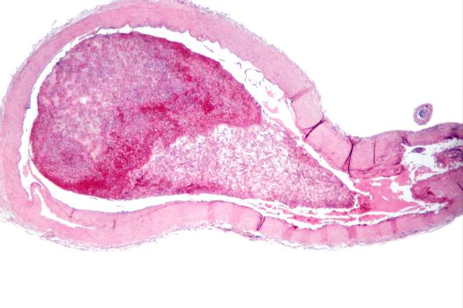
| 04:19, 21 August 2013 | IPLab10Mucor1.jpg (file) |  |
38 KB | Seung Park | This is a low-power photomicrograph of a section of carotid artery containing a mural thrombus. | 1 |
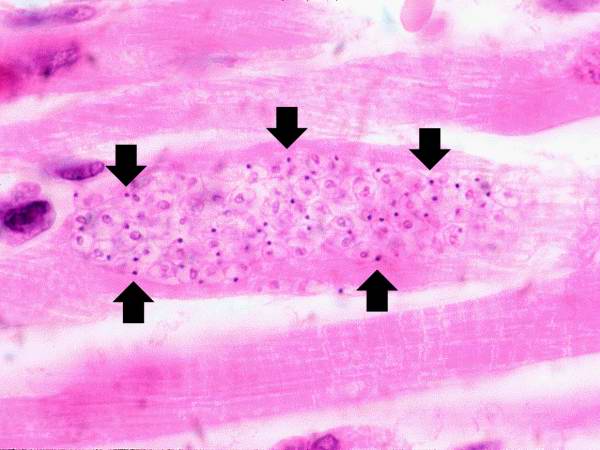
| 04:17, 21 August 2013 | IPLab10Blasto10.jpg (file) |  |
39 KB | Seung Park | This is a very high-power photomicrograph showing Blastomyces organisms stained with PAS. Note the budding organism (arrow) and the underlying pyogranulomatous inflammatory reaction in the background. | 1 |
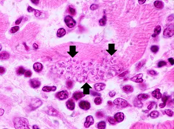
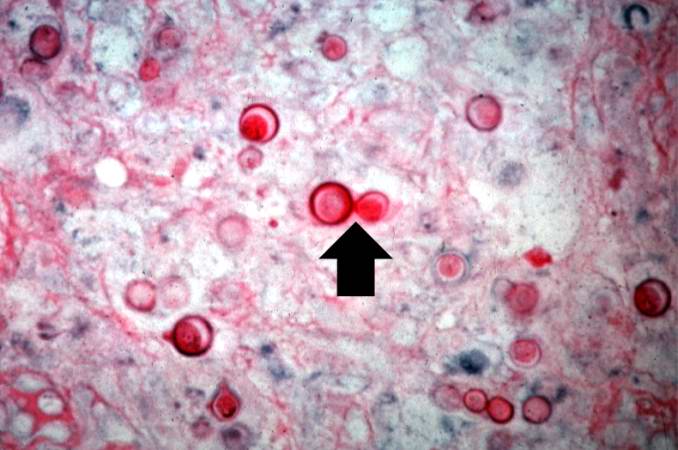
| 04:16, 21 August 2013 | IPLab10Blasto9.jpg (file) |  |
46 KB | Seung Park | This high-power photomicrograph shows Blastomyces organisms stained with PAS. Note the budding organism (arrow). Blastomyces has a characteristic presentation of budding which aids in diagnosis of the fungus. | 1 |
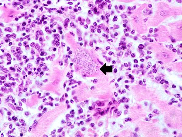
| 04:16, 21 August 2013 | IPLab10Blasto8.jpg (file) |  |
64 KB | Seung Park | This is a high-power photomicrograph showing an alveolus filled with Blastomyces organisms. This section is stained with Periodic Acid-Schiff (PAS) to stain the Blastomyces organisms. | 1 |
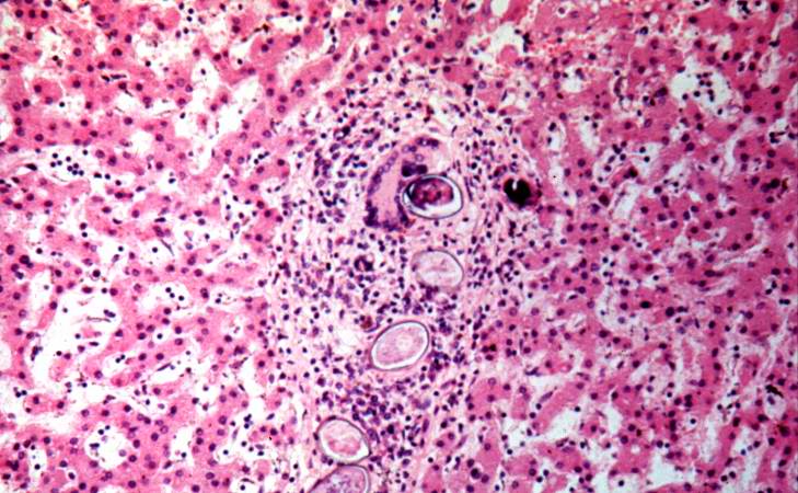
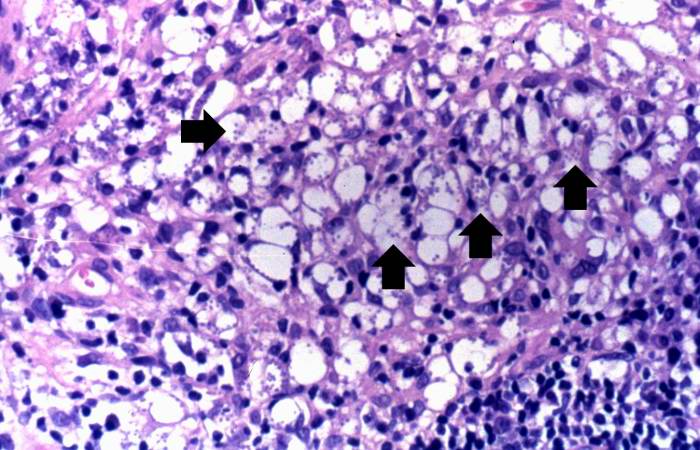
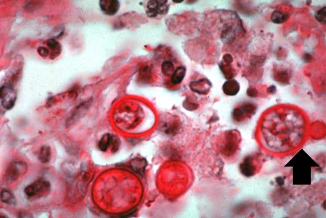
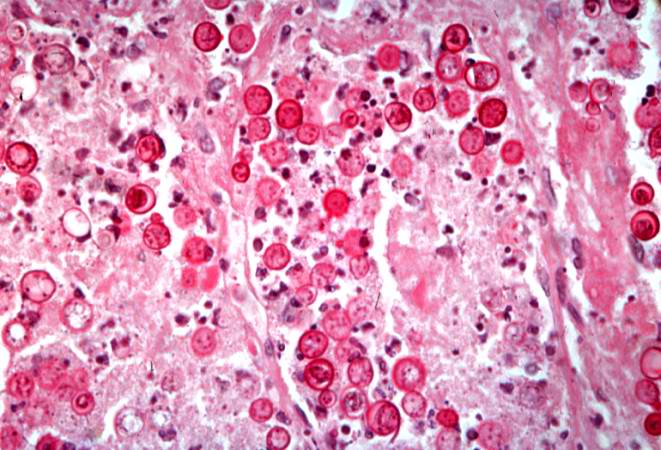
| 04:16, 21 August 2013 | IPLab10Blasto7.jpg (file) |  |
90 KB | Seung Park | This high-power photomicrograph shows an alveolus filled with numerous round bodies up to 25 mm in diameter. Some of these double-contour bodies (1) have a dense center and a clear halo. These are the Blastomyces organisms. The typical B. dermatitides ... | 1 |