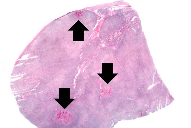
File:IPLab10Candidiasis3.jpg
Revision as of 04:03, 21 August 2013 by Seung Park (talk | contribs) (This is a low-power photomicrograph of lymph node with three prominent areas of Candida colonies (arrows). Even at this low magnification, the purple-staining yeast and pseudohyphae can be easily seen. This section was stained with Periodic Acid-Schiff...)
No higher resolution available.
IPLab10Candidiasis3.jpg (670 × 450 pixels, file size: 25 KB, MIME type: image/jpeg)
This is a low-power photomicrograph of lymph node with three prominent areas of Candida colonies (arrows). Even at this low magnification, the purple-staining yeast and pseudohyphae can be easily seen. This section was stained with Periodic Acid-Schiff Hematoxylin (PASH ), which stains the cell wall of fungi to make them more easily visible.
File history
Click on a date/time to view the file as it appeared at that time.
| Date/Time | Thumbnail | Dimensions | User | Comment | |
|---|---|---|---|---|---|
| current | 04:03, 21 August 2013 | | 670 × 450 (25 KB) | Seung Park (talk | contribs) | This is a low-power photomicrograph of lymph node with three prominent areas of Candida colonies (arrows). Even at this low magnification, the purple-staining yeast and pseudohyphae can be easily seen. This section was stained with Periodic Acid-Schiff... |
- You cannot overwrite this file.
File usage
The following page links to this file: