File:IPLab1Prostate3.jpg
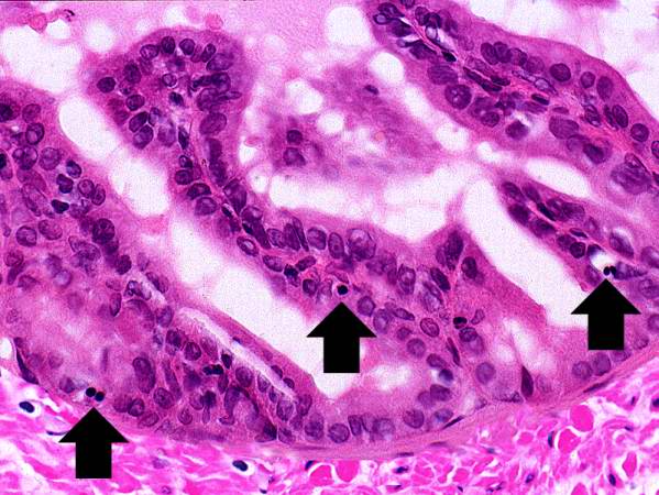
Revision as of 03:41, 16 August 2013 by Seung Park (talk | contribs) (Another high-power photomicrograph of the prostatic epithelium shows cells with pyknotic and fragmented nuclei (arrows). Note that the cytoplasm is condensed and hypereosinophilic.)
No higher resolution available.
IPLab1Prostate3.jpg (599 × 450 pixels, file size: 60 KB, MIME type: image/jpeg)
Another high-power photomicrograph of the prostatic epithelium shows cells with pyknotic and fragmented nuclei (arrows). Note that the cytoplasm is condensed and hypereosinophilic.
File history
Click on a date/time to view the file as it appeared at that time.
| Date/Time | Thumbnail | Dimensions | User | Comment | |
|---|---|---|---|---|---|
| current | 03:41, 16 August 2013 | | 599 × 450 (60 KB) | Seung Park (talk | contribs) | Another high-power photomicrograph of the prostatic epithelium shows cells with pyknotic and fragmented nuclei (arrows). Note that the cytoplasm is condensed and hypereosinophilic. |
- You cannot overwrite this file.
File usage
The following page links to this file: