File:IPLab3AcuteAppendicitis2.jpg
Revision as of 02:10, 19 August 2013 by Seung Park (talk | contribs) (This is a low-power photomicrograph of a normal appendix on the right and an appendix with acute inflammatory response on the left. Note the abundant blue-stained lymphoid tissue beneath the mucosal layer and the absence of blue-staining cells in the s...)
No higher resolution available.
IPLab3AcuteAppendicitis2.jpg (675 × 450 pixels, file size: 13 KB, MIME type: image/jpeg)
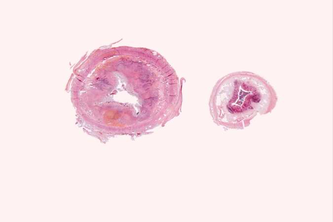
This is a low-power photomicrograph of a normal appendix on the right and an appendix with acute inflammatory response on the left. Note the abundant blue-stained lymphoid tissue beneath the mucosal layer and the absence of blue-staining cells in the submucosal layer of the normal appendix. Compare this with the extensive distribution of cells throughout the wall of the appendix with acute appendicitis. The blue color is due to the presence of many inflammatory cells, although at this low power these individual cells cannot be specifically identified.
File history
Click on a date/time to view the file as it appeared at that time.
| Date/Time | Thumbnail | Dimensions | User | Comment | |
|---|---|---|---|---|---|
| current | 02:10, 19 August 2013 | | 675 × 450 (13 KB) | Seung Park (talk | contribs) | This is a low-power photomicrograph of a normal appendix on the right and an appendix with acute inflammatory response on the left. Note the abundant blue-stained lymphoid tissue beneath the mucosal layer and the absence of blue-staining cells in the s... |
- You cannot overwrite this file.
File usage
The following page links to this file: