File:IPLab9Diphtheria3.jpg
Revision as of 03:50, 21 August 2013 by Seung Park (talk | contribs) (This is an even higher-power photomicrograph of the tracheal mucosa and the diphtheritic membrane. The mucosal surface of the trachea is ulcerated (total loss of epithelial cells) and the only remaining epithelial cells are found in the glands (arrows)...)
No higher resolution available.
IPLab9Diphtheria3.jpg (686 × 450 pixels, file size: 71 KB, MIME type: image/jpeg)
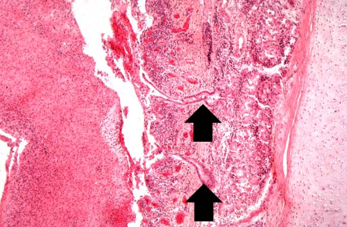
This is an even higher-power photomicrograph of the tracheal mucosa and the diphtheritic membrane. The mucosal surface of the trachea is ulcerated (total loss of epithelial cells) and the only remaining epithelial cells are found in the glands (arrows). The diphtheritic membrane consists of fibrin and inflammatory cells, most of which are dead.
File history
Click on a date/time to view the file as it appeared at that time.
| Date/Time | Thumbnail | Dimensions | User | Comment | |
|---|---|---|---|---|---|
| current | 03:50, 21 August 2013 | | 686 × 450 (71 KB) | Seung Park (talk | contribs) | This is an even higher-power photomicrograph of the tracheal mucosa and the diphtheritic membrane. The mucosal surface of the trachea is ulcerated (total loss of epithelial cells) and the only remaining epithelial cells are found in the glands (arrows)... |
- You cannot overwrite this file.
File usage
The following page links to this file: