File list
This special page shows all uploaded files.
| Date | Name | Thumbnail | Size | Description | Versions |
|---|---|---|---|---|---|
| 15:29, 19 August 2013 | IPLab2Hyperplasia9.jpg (file) |  |
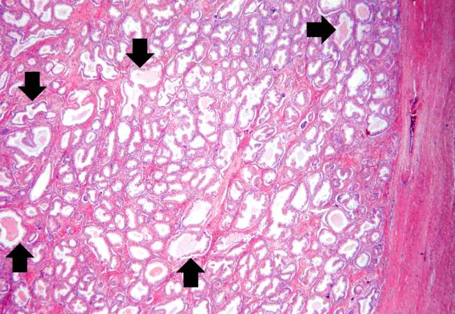
37 KB | This kidney was removed from another autopsy patient who had prostatic hyperplasia resulting in marked urinary retention and back-flow of urine from the bladder into the ureters and renal pelvis. The increased pressure inside the renal pelvis resulted ... | 1 |
| 15:29, 19 August 2013 | IPLab2Hyperplasia8.jpg (file) |  |
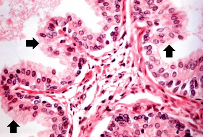
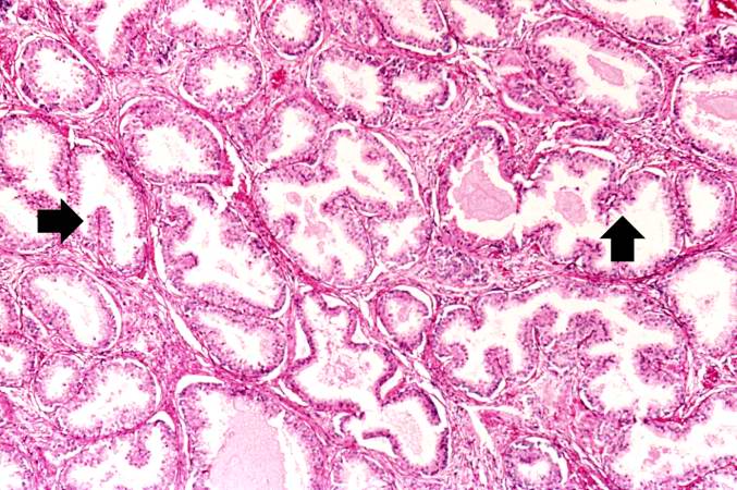
53 KB | This is a higher-power photomicrograph of papillary folds of hyperplastic epithelium (arrows). | 1 |
| 15:29, 19 August 2013 | IPLab2Hyperplasia7.jpg (file) |  |
74 KB | A higher-power view shows the papillary folds (arrows) produced by the hyperplastic epithelium projecting into the lumen of the gland. While these papillary folds project into the lumen of the gland, there is no extension through the glandular basement... | 1 |
| 15:28, 19 August 2013 | IPLab2Hyperplasia6.jpg (file) |  |
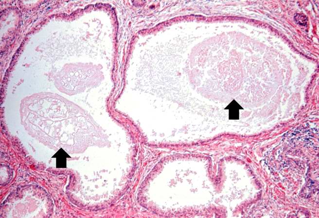
61 KB | Cystic dilatation of glands is present in this photomicrograph. Notice the accumulation of secretory material inside the glands (arrows) and compression (thinning) of the lining epithelium. | 1 |
| 15:28, 19 August 2013 | IPLab2Hyperplasia5.jpg (file) |  |
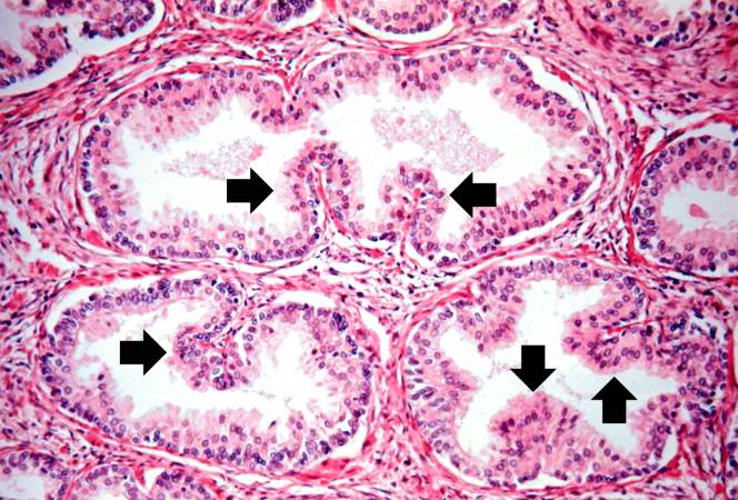
86 KB | Note these glands, which exhibit hyperplasia of the glandular epithelium. The infolding of the glandular epithelial cells forms papillary projections (arrows) into the lumen of the gland. | 1 |
| 15:28, 19 August 2013 | IPLab2Hyperplasia4.jpg (file) |  |
69 KB | The dilated glands (arrows) make up the major portion of the prostate tissue and there is compression of the stroma. | 1 |
| 15:28, 19 August 2013 | IPLab2Hyperplasia3.jpg (file) |  |
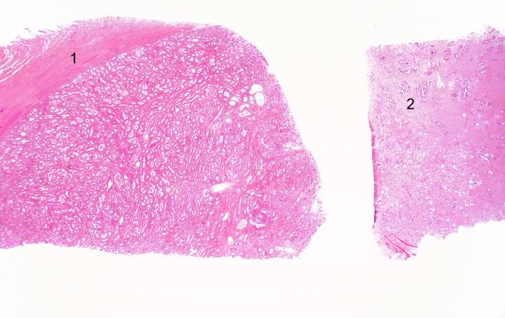
48 KB | This is a low-power photomicrograph showing hyperplastic prostate on the left (1) and normal prostate on the right (2). At this power, dilated glands are visible in the section of hyperplastic prostate. | 1 |
| 15:27, 19 August 2013 | IPLab2Hyperplasia2.jpg (file) |  |
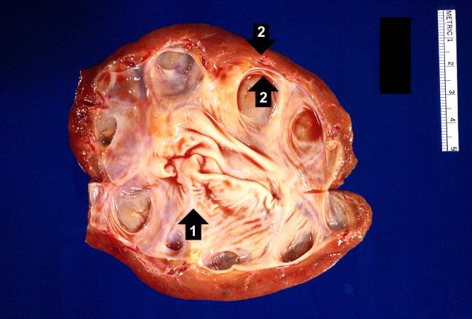
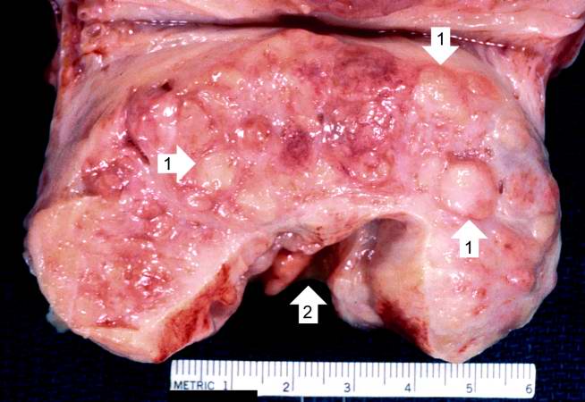
44 KB | This is a close-up of the prostate from this same patient. Note the nodularity of the tissue (1) and the enlargement of the gland. Enlargement of the prostate leads to compression of the urethra as it passes through (2) the gland. | 1 |
| 15:27, 19 August 2013 | IPLab2Hyperplasia1.jpg (file) |  |
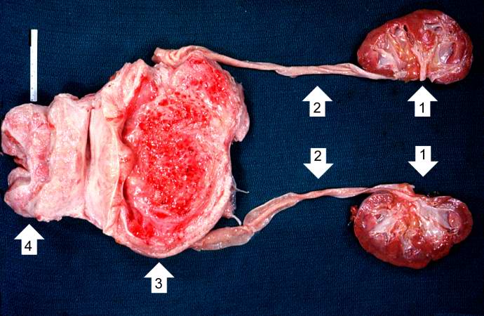
58 KB | This photograph shows the autopsy specimen from this patient. Included are kidneys (1), ureters (2), bladder (3) (which has been opened), and enlarged prostate (4). Note that the bladder mucosa has multiple trabeculae and the bladder mucosa is hyperemi... | 1 |
| 14:25, 14 August 2013 | Test.jpg (file) |  |
270 KB | This is a test of the uploading system. | 1 |