No higher resolution available.
IPLab1LungAbscess1.jpg (307 × 450 pixels, file size: 30 KB, MIME type: image/jpeg)
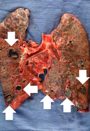
This is a gross photograph of the lungs from this case. Note the abscesses (arrows) especially in the lower lobes. The entire lung is consolidated.
An abscess is a collection of pus (white blood cells) within a cavity formed by disintegrated tissue.
Consolidation is the filling of lung air spaces with exudate--this is a sign of pneumonia.
File history
Click on a date/time to view the file as it appeared at that time.
| Date/Time | Thumbnail | Dimensions | User | Comment | |
|---|---|---|---|---|---|
| current | 16:14, 15 August 2013 | | 307 × 450 (30 KB) | Seung Park (talk | contribs) |
- You cannot overwrite this file.
File usage
The following page links to this file: