File:IPLab1Tuberculosis1.jpg
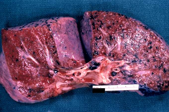
This is a gross photograph of a cut section of lung from this patient with disseminated tuberculosis. The numerous small white nodules scattered throughout this lung tissue represent individual tuberculosis granulomas. In addition, note the dark areas throughout the lung which represent deposits of anthracotic pigment.
Disseminated tuberculosis refers to the hematogenous spread of tuberculous lesions throughout the body. It is also known as miliary tuberculosis (which is so-called because the lesions resemble millet).
A tuberculosis granuloma is a focus of granulomatous inflammation caused by CHRONIC tuberculosis infection. The granuloma consists of epithelioid cells (activated macrophages) surrounded by lymphocytes, plasma cells, and fibroblasts.
Anthracotic pigment is coal dust deposited in the lungs--it is seen in coal miners, city-dwellers, and smokers.
File history
Click on a date/time to view the file as it appeared at that time.
| Date/Time | Thumbnail | Dimensions | User | Comment | |
|---|---|---|---|---|---|
| current | 02:49, 16 August 2013 | | 680 × 450 (66 KB) | Seung Park (talk | contribs) |
- You cannot overwrite this file.
File usage
The following page links to this file: