Clinical Summary[edit]
This white female infant was the product of an uncomplicated term delivery, though meconium staining was noted at birth. During the first day post-partum, the infant's abdomen became progressively distended and a meconium ileus was suspected. Surgery confirmed the presence of a meconium ileus and a section of perforated atretic jejunum proximal to the ileus was resected. Eight days later, the patient's condition had deteriorated. A second operation revealed a segment of necrotic bowel, which was removed. Subsequently the infant's pulmonary function deteriorated and she required frequent suctioning. She developed repeated episodes of pneumonia (E. coli and Pseudomonas grew out on cultures) complicated by atelectasis secondary to pneumothorax. The patient died at 25-days-of-age in respiratory failure.
Autopsy Findings[edit]
Bilateral, extensive organizing bronchopneumonia was present with evidence of a pneumothorax and atelectasis. There were significant changes in the pancreas consistent with cystic fibrosis as well as involvement of the small intestine and changes related to the surgical procedures.
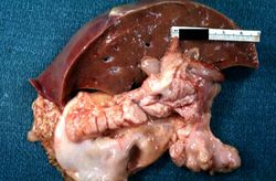
This is a gross photograph of liver and pancreas from the autopsy. The pancreas is slightly smaller than normal and it has a mucous consistency.
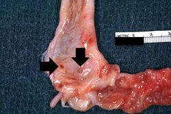
This section of duodenum demonstrates dilation, loss of rugae, and areas of ulceration (arrows).
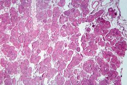
This low-power photomicrograph of pancreas shows increased interstitial connective tissue resulting in accentuation of the lobular pattern.
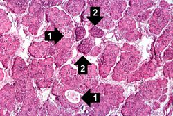
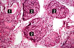
This higher-power photomicrograph of the pancreas shows interstitial tissue and the presence of small cystic spaces (1) within the acinar lobules. These spaces are filled with an eosinophilic proteinaceous material. The islets of Langerhans (2) are unaffected.
This higher-power photomicrograph shows a cystic space (1) within an acinar lobule. Islets of Langerhans (2) are also visible.
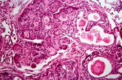
This high-power photomicrograph shows more clearly these variably-sized cystic spaces within the acinar pancreas.
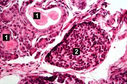
This is another high-power photomicrograph showing cystic spaces (1) within the acinar pancreas and a normal islet of Langerhans (2).
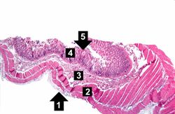
This low-power photomicrograph of intestine shows the normal layers of the intestine, including the serosa (1), the muscularis (2), the submucosa (3), and the mucosal layer (4) with its deep mucosal crypts. There is yet another cystic space within the mucosa (5).
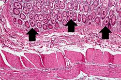
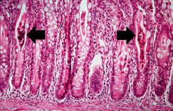
A higher-power photomicrograph shows the bottom of the intestinal crypts and the other normal layers of the intestine. Even at this magnification, accumulations of eosinophilic debris can be seen in many of the intestinal crypts (arrows).
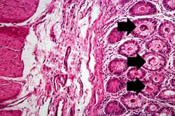
This is a higher-power photomicrograph showing the eosinophilic debris in many of the intestinal crypts (arrows).
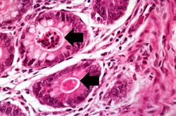
This higher-power photomicrograph shows more clearly the eosinophilic debris (arrows) in the intestinal crypts.
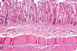
This is a low-power photomicrograph from another section of the intestine. Saggital sections of the intestinal crypts show the crypts along their full length, extending to the mucosal surface.
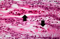
A higher-power photomicrograph of intestine shows the vacuolated intestinal epithelial cells lining the crypts and necrotic debris and inspissated secretions within the crypts (arrows).
Another high-power photomicrograph of intestine shows the vacuolated intestinal epithelial cells lining the crypts and necrotic debris and inspissated secretions within the crypts (arrows).
Study Questions[edit]
CF is autosomal recessive.
The frequency of CF in whites is 1/200. Thus, one out of every 20 whites must be heterozygous (heterozygotes have no recognizable symptoms).
Other racial groups have a very low incidence of CF.
CF causes altered cellular chloride transport leading to increased chloride levels in the sweat.
A sweat chloride level greater than 60 mEq/L is diagnostic for CF if other clinical signs are also present.
When children are 2-12 months of age they develop malodorous steatorrhea and recurrent chronic pulmonary infections. Lower than normal weight gain is also common.
Lung - chronic pneumonia, viscous mucous secretions, distended bronchioles, hypertrophy and hyperplasia of mucus-secreting glands, chronic bronchitis and bronchiolitis.
Pancreas - atrophy of exocrine pancreas, plugging of ducts, progressive fibrosis and fatty replacement. Insufficiency of exocrine pancreas leads to steatorrhea and fat soluble vitamin deficiencies (e.g., low vitamin K leads to bleeding disorders).
Liver - plugging of bile canaliculi, biliary cirrhosis.
Salivary glands - dilation of ducts, plugging, squamous metaplasia of lining epithelium, fibrosis.
Wolffian duct obstruction - infertility in males who live to puberty.
Additional Resources[edit]
Reference[edit]
Journal Articles[edit]
Related IPLab Cases[edit]
