Cytologically Yours: CoW: 20131216
Revision as of 22:05, 14 January 2014 by Stephanie Simmons (talk | contribs)
Contents
Clinical Summary
The patient is an 66 year old white male with a history of smoking, COPD, and diabetes. The patient presented with increased shortness of breath.
Past Medical History
- Diabetes
- COPD
- Squamous cell carcinoma of skin
Past Surgical History
- Excision of squamous cell carcinoma
- Removal of adenomatous polyp of sigmoid colon
Clinical Plan
The differential diagnosis includes worsening of COPD. CT imaging of chest is performed.
Radiology
- CT Chest shows hilar lung mass and multiple mediastinal lymph nodes showing increased uptake on PET scan.
Pathology
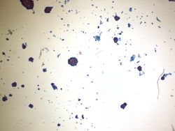
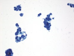
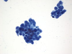
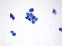
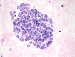
Cytology
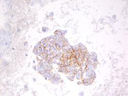
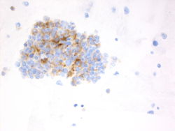
Immunohistochemistry
Other immunostains performed
- BerEp4 Positive
- Moc31 Faintly positive
- Calretinin Negative
- TTF1 Negative
- Chromogranin Positive
- Synaptophysin Positive
- CD56 Positive
- Napsin A Negative
Resident Questions
- <spoiler text="
</spoiler>
Final Diagnosis
Cytology
- Small cell carcinoma
Case Discussion
This is a classic case of metastatic small cell carcinoma in pleural fluid.
| ||||||||
