File:IPLab3AcuteAppendicitis4.jpg
Revision as of 02:11, 19 August 2013 by Seung Park (talk | contribs) (This is a photomicrograph of the serosal surface of the appendix on the left (1) and the submucosal tissue in the center (2) with remnants of a lymphoid nodule. Surrounding this lymphoid nodule are masses of leukocytes which should not be present in a ...)
No higher resolution available.
IPLab3AcuteAppendicitis4.jpg (681 × 450 pixels, file size: 71 KB, MIME type: image/jpeg)
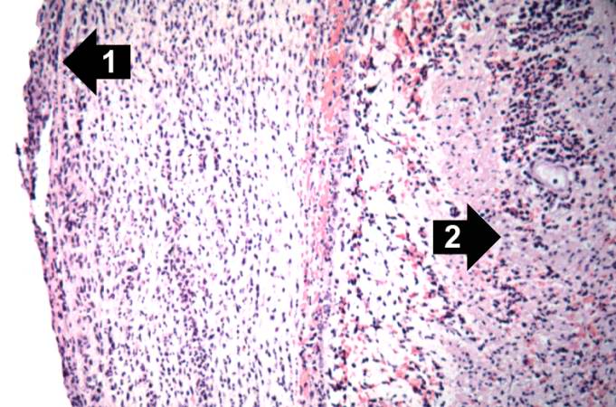
This is a photomicrograph of the serosal surface of the appendix on the left (1) and the submucosal tissue in the center (2) with remnants of a lymphoid nodule. Surrounding this lymphoid nodule are masses of leukocytes which should not be present in a normal appendix.
File history
Click on a date/time to view the file as it appeared at that time.
| Date/Time | Thumbnail | Dimensions | User | Comment | |
|---|---|---|---|---|---|
| current | 02:11, 19 August 2013 | | 681 × 450 (71 KB) | Seung Park (talk | contribs) | This is a photomicrograph of the serosal surface of the appendix on the left (1) and the submucosal tissue in the center (2) with remnants of a lymphoid nodule. Surrounding this lymphoid nodule are masses of leukocytes which should not be present in a ... |
- You cannot overwrite this file.
File usage
The following page links to this file: