File:IPLab3AcuteAppendicitis6.jpg
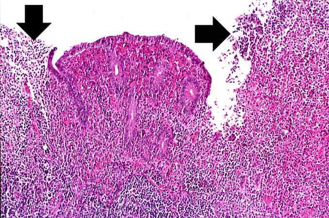
Revision as of 02:12, 19 August 2013 by Seung Park (talk | contribs) (This higher-power photomicrograph of the mucosal surface shows the loss of normal mucosal epithelium (arrows) and the inflammatory infiltrate. The principal inflammatory cell in this case of acute appendicitis is the neutrophil.)
No higher resolution available.
IPLab3AcuteAppendicitis6.jpg (678 × 450 pixels, file size: 106 KB, MIME type: image/jpeg)
This higher-power photomicrograph of the mucosal surface shows the loss of normal mucosal epithelium (arrows) and the inflammatory infiltrate. The principal inflammatory cell in this case of acute appendicitis is the neutrophil.
An infiltrate is an accumulation of cells in the lung parenchyma--this is a sign of pneumonia.
File history
Click on a date/time to view the file as it appeared at that time.
| Date/Time | Thumbnail | Dimensions | User | Comment | |
|---|---|---|---|---|---|
| current | 02:12, 19 August 2013 | | 678 × 450 (106 KB) | Seung Park (talk | contribs) | This higher-power photomicrograph of the mucosal surface shows the loss of normal mucosal epithelium (arrows) and the inflammatory infiltrate. The principal inflammatory cell in this case of acute appendicitis is the neutrophil. |
- You cannot overwrite this file.
File usage
The following page links to this file: