File:IPLab3LobarPneumonia6.jpg
Revision as of 03:18, 19 August 2013 by Seung Park (talk | contribs) (This photomicrograph of the lung shows the junction of the pleura (1) with the pneumonic lung parenchyma (2). Careful examination of the lung tissue at the right reveals the outline of alveolar structures in this tissue (arrows). The mass of cells infi...)
No higher resolution available.
IPLab3LobarPneumonia6.jpg (664 × 450 pixels, file size: 80 KB, MIME type: image/jpeg)
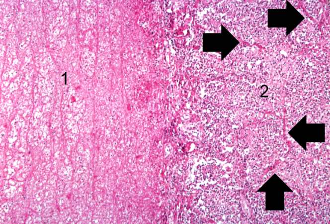
This photomicrograph of the lung shows the junction of the pleura (1) with the pneumonic lung parenchyma (2). Careful examination of the lung tissue at the right reveals the outline of alveolar structures in this tissue (arrows). The mass of cells infiltrating into this lung tissue consists almost exclusively of polymorphonuclear leukocytes (neutrophils).
File history
Click on a date/time to view the file as it appeared at that time.
| Date/Time | Thumbnail | Dimensions | User | Comment | |
|---|---|---|---|---|---|
| current | 03:18, 19 August 2013 | | 664 × 450 (80 KB) | Seung Park (talk | contribs) | This photomicrograph of the lung shows the junction of the pleura (1) with the pneumonic lung parenchyma (2). Careful examination of the lung tissue at the right reveals the outline of alveolar structures in this tissue (arrows). The mass of cells infi... |
- You cannot overwrite this file.
File usage
The following page links to this file: