File:IPLab3ChronicPepticUlcer3.jpg
Revision as of 04:15, 19 August 2013 by Seung Park (talk | contribs) (This is a low-power photomicrograph of the transected ulcer. The blue cells on the right hand side of this section are the normal gastric epithelial cells of the mucosa (1). Note the absence of any epithelial cells within the crater of the ulcer (2).)
No higher resolution available.
IPLab3ChronicPepticUlcer3.jpg (686 × 450 pixels, file size: 31 KB, MIME type: image/jpeg)
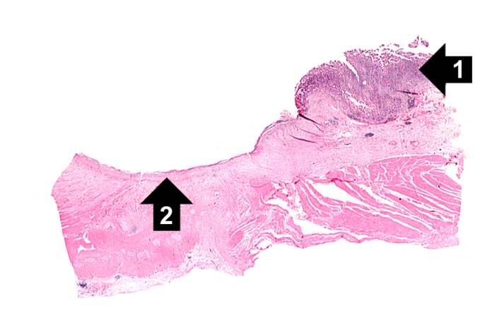
This is a low-power photomicrograph of the transected ulcer. The blue cells on the right hand side of this section are the normal gastric epithelial cells of the mucosa (1). Note the absence of any epithelial cells within the crater of the ulcer (2).
File history
Click on a date/time to view the file as it appeared at that time.
| Date/Time | Thumbnail | Dimensions | User | Comment | |
|---|---|---|---|---|---|
| current | 04:15, 19 August 2013 | | 686 × 450 (31 KB) | Seung Park (talk | contribs) | This is a low-power photomicrograph of the transected ulcer. The blue cells on the right hand side of this section are the normal gastric epithelial cells of the mucosa (1). Note the absence of any epithelial cells within the crater of the ulcer (2). |
- You cannot overwrite this file.
File usage
The following page links to this file: