File:IPLab4Thrombosis6.jpg
Revision as of 16:39, 19 August 2013 by Seung Park (talk | contribs) (This is a higher-power photomicrograph of thrombus attached to the wall of the vessel. Note the early organization with in-growth of fibroblasts and small blood vessels from the wall of the artery (arrows).)
No higher resolution available.
IPLab4Thrombosis6.jpg (682 × 450 pixels, file size: 66 KB, MIME type: image/jpeg)
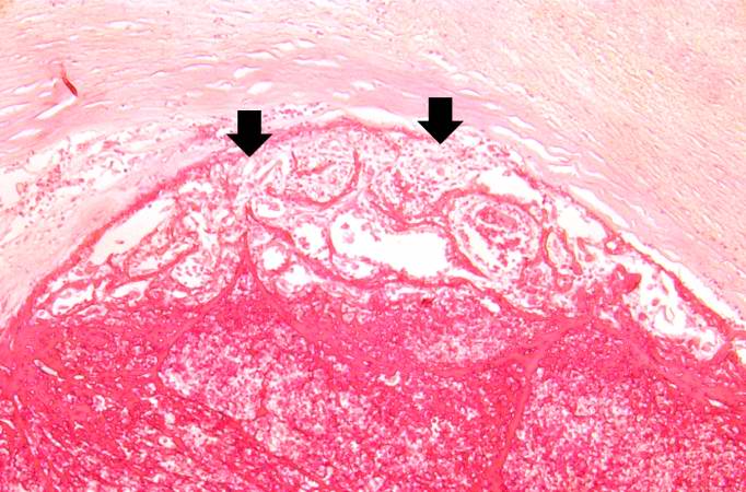
This is a higher-power photomicrograph of thrombus attached to the wall of the vessel. Note the early organization with in-growth of fibroblasts and small blood vessels from the wall of the artery (arrows).
A thrombus is a solid mass resulting from the aggregation of blood constituents within the vascular system.
File history
Click on a date/time to view the file as it appeared at that time.
| Date/Time | Thumbnail | Dimensions | User | Comment | |
|---|---|---|---|---|---|
| current | 16:39, 19 August 2013 | | 682 × 450 (66 KB) | Seung Park (talk | contribs) | This is a higher-power photomicrograph of thrombus attached to the wall of the vessel. Note the early organization with in-growth of fibroblasts and small blood vessels from the wall of the artery (arrows). |
- You cannot overwrite this file.
File usage
The following page links to this file: