File:IPLab6AcuteRejection7.jpg
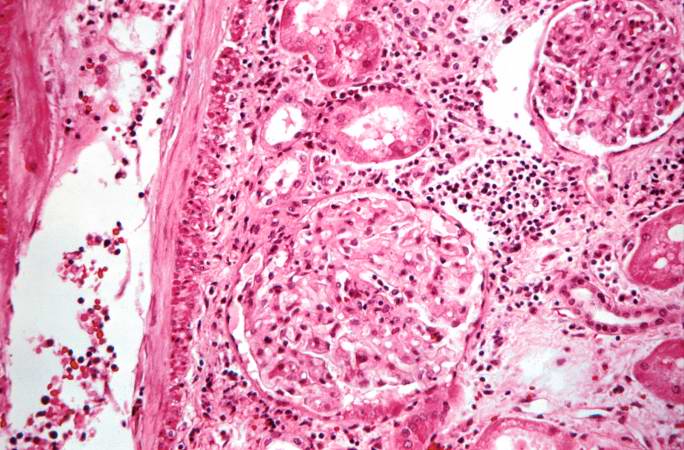
Revision as of 21:57, 20 August 2013 by Seung Park (talk | contribs) (This high-power photomicrograph demonstrates the cellular infiltrate within the interstitium and in the wall of the blood vessel on the left.)
No higher resolution available.
IPLab6AcuteRejection7.jpg (684 × 450 pixels, file size: 74 KB, MIME type: image/jpeg)
This high-power photomicrograph demonstrates the cellular infiltrate within the interstitium and in the wall of the blood vessel on the left.
An infiltrate is an accumulation of cells in the lung parenchyma--this is a sign of pneumonia.
File history
Click on a date/time to view the file as it appeared at that time.
| Date/Time | Thumbnail | Dimensions | User | Comment | |
|---|---|---|---|---|---|
| current | 21:57, 20 August 2013 | | 684 × 450 (74 KB) | Seung Park (talk | contribs) | This high-power photomicrograph demonstrates the cellular infiltrate within the interstitium and in the wall of the blood vessel on the left. |
- You cannot overwrite this file.
File usage
The following page links to this file: