File:IPLab7Adenoma3.jpg
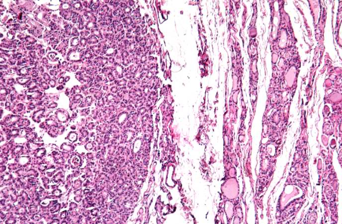
Revision as of 01:22, 21 August 2013 by Seung Park (talk | contribs) (This is another higher-power photomicrograph of the adenoma (left) and the adjacent thyroid tissue (right). Note the compression of the adjacent normal thyroid and the difference in morphology between the adenoma and the thyroid.)
No higher resolution available.
IPLab7Adenoma3.jpg (688 × 450 pixels, file size: 89 KB, MIME type: image/jpeg)
This is another higher-power photomicrograph of the adenoma (left) and the adjacent thyroid tissue (right). Note the compression of the adjacent normal thyroid and the difference in morphology between the adenoma and the thyroid.
File history
Click on a date/time to view the file as it appeared at that time.
| Date/Time | Thumbnail | Dimensions | User | Comment | |
|---|---|---|---|---|---|
| current | 01:22, 21 August 2013 | | 688 × 450 (89 KB) | Seung Park (talk | contribs) | This is another higher-power photomicrograph of the adenoma (left) and the adjacent thyroid tissue (right). Note the compression of the adjacent normal thyroid and the difference in morphology between the adenoma and the thyroid. |
- You cannot overwrite this file.
File usage
The following page links to this file: