File:IPLab7LipSCC6.jpg
Revision as of 01:32, 21 August 2013 by Seung Park (talk | contribs) (This is a high power photomicrograph of the well-differentiated squamous cell carcinoma. Note the intracytoplasmic keratinization which gives the cells a glassy appearance. The focal accumulations of keratinized cells are called keratin pearls (arrows).)
No higher resolution available.
IPLab7LipSCC6.jpg (671 × 450 pixels, file size: 85 KB, MIME type: image/jpeg)
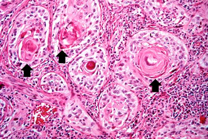
This is a high power photomicrograph of the well-differentiated squamous cell carcinoma. Note the intracytoplasmic keratinization which gives the cells a glassy appearance. The focal accumulations of keratinized cells are called keratin pearls (arrows).
File history
Click on a date/time to view the file as it appeared at that time.
| Date/Time | Thumbnail | Dimensions | User | Comment | |
|---|---|---|---|---|---|
| current | 01:32, 21 August 2013 | | 671 × 450 (85 KB) | Seung Park (talk | contribs) | This is a high power photomicrograph of the well-differentiated squamous cell carcinoma. Note the intracytoplasmic keratinization which gives the cells a glassy appearance. The focal accumulations of keratinized cells are called keratin pearls (arrows). |
- You cannot overwrite this file.
File usage
The following page links to this file: