File:IPLab7Metastatic9.jpg
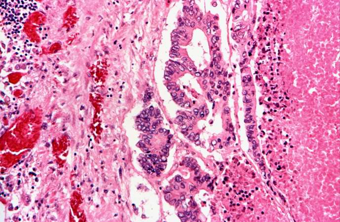
Revision as of 01:47, 21 August 2013 by Seung Park (talk | contribs) (This is a high-power photomicrograph of the edge of the tumor nodule in the lung. The tumor cells area growing in a glandular pattern. The area of necrosis is evident at the right side of the image.)
No higher resolution available.
IPLab7Metastatic9.jpg (689 × 450 pixels, file size: 78 KB, MIME type: image/jpeg)
This is a high-power photomicrograph of the edge of the tumor nodule in the lung. The tumor cells area growing in a glandular pattern. The area of necrosis is evident at the right side of the image.
File history
Click on a date/time to view the file as it appeared at that time.
| Date/Time | Thumbnail | Dimensions | User | Comment | |
|---|---|---|---|---|---|
| current | 01:47, 21 August 2013 | | 689 × 450 (78 KB) | Seung Park (talk | contribs) | This is a high-power photomicrograph of the edge of the tumor nodule in the lung. The tumor cells area growing in a glandular pattern. The area of necrosis is evident at the right side of the image. |
- You cannot overwrite this file.
File usage
The following page links to this file: