File:IPLab7IDC2.jpg
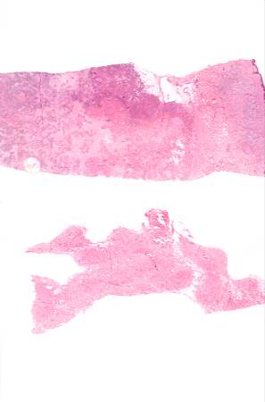
Revision as of 01:50, 21 August 2013 by Seung Park (talk | contribs) (These are sections of normal breast (lower) and breast tissue with infiltrating duct carcinoma (upper). Note the increased cellularity (increased blue staining due to the increased number of nuclei) in the tumor tissue.)
No higher resolution available.
IPLab7IDC2.jpg (297 × 450 pixels, file size: 12 KB, MIME type: image/jpeg)
These are sections of normal breast (lower) and breast tissue with infiltrating duct carcinoma (upper). Note the increased cellularity (increased blue staining due to the increased number of nuclei) in the tumor tissue.
File history
Click on a date/time to view the file as it appeared at that time.
| Date/Time | Thumbnail | Dimensions | User | Comment | |
|---|---|---|---|---|---|
| current | 01:50, 21 August 2013 | | 297 × 450 (12 KB) | Seung Park (talk | contribs) | These are sections of normal breast (lower) and breast tissue with infiltrating duct carcinoma (upper). Note the increased cellularity (increased blue staining due to the increased number of nuclei) in the tumor tissue. |
- You cannot overwrite this file.
File usage
The following page links to this file: