File:IPLab7IDC8.jpg
Revision as of 01:51, 21 August 2013 by Seung Park (talk | contribs) (This is a high-power photomicrograph demonstrating the growth pattern of the tumor. The tumor consists of malignant duct-lining cells growing in cords, solid cell nests, tubules, and glands. The cytologic detail of tumor cells varies from small cells w...)
No higher resolution available.
IPLab7IDC8.jpg (691 × 450 pixels, file size: 73 KB, MIME type: image/jpeg)
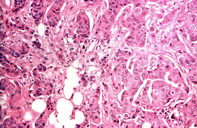
This is a high-power photomicrograph demonstrating the growth pattern of the tumor. The tumor consists of malignant duct-lining cells growing in cords, solid cell nests, tubules, and glands. The cytologic detail of tumor cells varies from small cells with moderately hyperchromatic, regular nuclei to large cells with large, irregular, hyperchromatic nuclei.
File history
Click on a date/time to view the file as it appeared at that time.
| Date/Time | Thumbnail | Dimensions | User | Comment | |
|---|---|---|---|---|---|
| current | 01:51, 21 August 2013 | | 691 × 450 (73 KB) | Seung Park (talk | contribs) | This is a high-power photomicrograph demonstrating the growth pattern of the tumor. The tumor consists of malignant duct-lining cells growing in cords, solid cell nests, tubules, and glands. The cytologic detail of tumor cells varies from small cells w... |
- You cannot overwrite this file.
File usage
The following page links to this file: