From Pathology Education Instructional Resource
Images
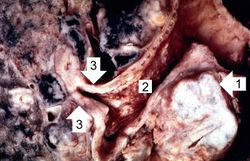
This is a gross photograph of bronchogenic carcinoma. The large tumor mass can be seen adjacent to the bronchus (1). Note that the epithelial surface of the bronchus is rough and irregular (2). The first branch off the right main stem bronchus is partially occluded by the thickened mucosa and submucosa (3).
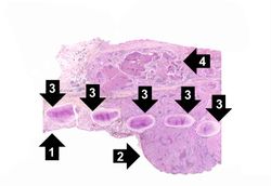
This is a low-power photomicrograph of bronchus showing normal mucosa (1) with transition to carcinoma (2). Note the bronchial cartilage (3) and the invasion of tumor through the entire wall of the bronchus with tumor extending to the serosal surface (4).
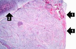
This is a photomicrograph of bronchus with ulcerated mucosal surface on the right (1). The submucosa is completely filled with tumor down to the cartilage (2).
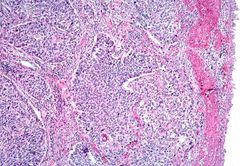
This is a higher-power photomicrograph of bronchus with the ulcerated mucosal surface on the right and tumor underneath.
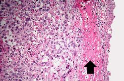
This is a higher-power photomicrograph of the mucosal surface (right) with an area of hemorrhage (arrow) and underlying tumor (left).
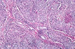
This is a photomicrograph of tumor from an area of invasion with compression of fibrous stroma and focal necrosis.
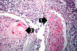
This is a high-power photomicrograph showing cytologic detail of the tumor with an area of necrosis (1) and a more differentiated area with keratin pearl formation (2).
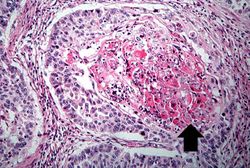
This is a high power photomicrograph of tumor with an area of central necrosis (arrow).
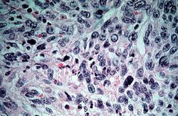
This high-power photomicrograph of tumor shows the cytologic detail of a less-differentiated area of neoplasm with cellular anaplasia.
