File:IPLab8HSVGlossitis5.jpg
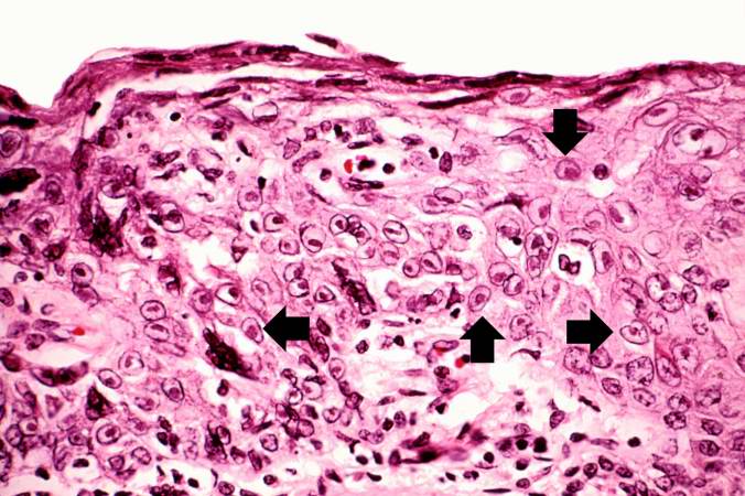
Revision as of 02:29, 21 August 2013 by Seung Park (talk | contribs) (This is a high-power photomicrograph of epithelium near the edge of the ulcer. The cells that have been invaded by the herpes virus contain intranuclear accumulations of amphophilic viral inclusions (arrows).)
No higher resolution available.
IPLab8HSVGlossitis5.jpg (676 × 450 pixels, file size: 70 KB, MIME type: image/jpeg)
This is a high-power photomicrograph of epithelium near the edge of the ulcer. The cells that have been invaded by the herpes virus contain intranuclear accumulations of amphophilic viral inclusions (arrows).
File history
Click on a date/time to view the file as it appeared at that time.
| Date/Time | Thumbnail | Dimensions | User | Comment | |
|---|---|---|---|---|---|
| current | 02:29, 21 August 2013 | | 676 × 450 (70 KB) | Seung Park (talk | contribs) | This is a high-power photomicrograph of epithelium near the edge of the ulcer. The cells that have been invaded by the herpes virus contain intranuclear accumulations of amphophilic viral inclusions (arrows). |
- You cannot overwrite this file.
File usage
The following page links to this file: