File:IPLab8HSVEncephalitis4.jpg
Revision as of 02:35, 21 August 2013 by Seung Park (talk | contribs) (This is a medium-power photomicrograph showing a blood vessel with perivascular hemorrhage (1), areas with loss of brain parenchyma, and edema (2). Even at this power, it can be seen that many of the cells are shrunken and dark red, suggesting that the...)
No higher resolution available.
IPLab8HSVEncephalitis4.jpg (670 × 450 pixels, file size: 58 KB, MIME type: image/jpeg)
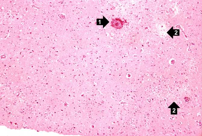
This is a medium-power photomicrograph showing a blood vessel with perivascular hemorrhage (1), areas with loss of brain parenchyma, and edema (2). Even at this power, it can be seen that many of the cells are shrunken and dark red, suggesting that they are necrotic.
File history
Click on a date/time to view the file as it appeared at that time.
| Date/Time | Thumbnail | Dimensions | User | Comment | |
|---|---|---|---|---|---|
| current | 02:35, 21 August 2013 | | 670 × 450 (58 KB) | Seung Park (talk | contribs) | This is a medium-power photomicrograph showing a blood vessel with perivascular hemorrhage (1), areas with loss of brain parenchyma, and edema (2). Even at this power, it can be seen that many of the cells are shrunken and dark red, suggesting that the... |
- You cannot overwrite this file.
File usage
The following page links to this file: