From Pathology Education Instructional Resource
Images
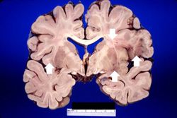
This is a gross photograph of a section of brain showing multiple small, punctate hemorrhages throughout the brain parenchyma (arrows).
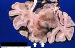
This is a closer view of the previous section of brain showing multiple small, punctate hemorrhages throughout the brain parenchyma (arrows).
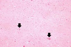
This is a low-power photomicrograph showing a section of brain with numerous perivascular hemorrhages (arrows) and some areas that appear hypercellular.
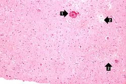
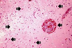
This is a medium-power photomicrograph showing a blood vessel with perivascular hemorrhage (1), areas with loss of brain parenchyma, and edema (2). Even at this power, it can be seen that many of the cells are shrunken and dark red, suggesting that they are necrotic.
This is a high-power photomicrograph of the previous section. At this power it is easier to see the blood vessel with the perivascular hemorrhage and mild perivascular lymphocytic cuffing (1). In addition, the areas of edema and loss of neurophil (2) can be better appreciated. Red shrunken neurons and glia with pyknotic nuclei (3) are also evident at this power.
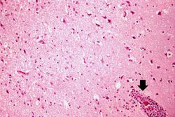
This is another high-power photomicrograph showing a blood vessel with perivascular hemorrhage and mild perivascular lymphocytic cuffing (arrow). In addition, there are numerous red shrunken neurons and glia with pyknotic nuclei throughout this section.
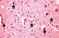
This is a high-power photomicrograph demonstrating clear areas, which indicate edema, and numerous shrunken red necrotic cells (1). At this power, it can be seen that eosinophilic intranuclear inclusion bodies have displaced chromatin to the periphery of the nucleus in some cells (2).
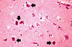
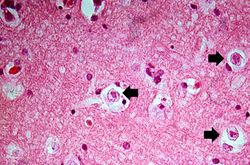
This is a high-power photomicrograph showing several necrotic cells (arrows).
This is a high-power photomicrograph demonstrating cells containing intranuclear inclusion bodies (arrows).
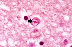
This is another high-power photomicrograph of a cell containing an intranuclear inclusion body (arrow). Note that the nuclear chromatin has been pushed to the outer edges of the nucleus.
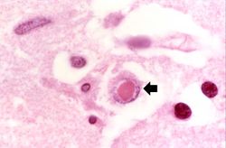
This is another high-power photomicrograph of a cell containing an intranuclear inclusion body (arrow).
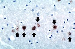
This is a photomicrograph of a brain section stained with an antibody against herpes simplex. Even at this magnification, it is easy to pick out cells that are positive for the virus (arrows).
